Common Skin Cell Types and Their Roles
The skin's most common cells build barriers, while rarer cells handle color, immunity, and touch.

Keratinocytes and How They Build the Skin's Physical Barrier
Keratinocytes are the epidermis. More than ninety percent of epidermal cells are keratinocytes, and their defining product, keratin, is a structural protein tough enough to resist heat, UV radiation, water loss, bacteria, fungi, and viruses simultaneously. The barrier isn't passive. It performs.
What I find fascinating about keratinocytes, and what took me a while to fully appreciate, is that the barrier isn't a thing that exists so much as a thing that's constantly being made. Keratinocytes are born in the stratum basale and spend their entire lives moving upward through biochemically distinct stages, shedding their nuclei, flattening into corneocytes, and stacking ten to thirty layers deep in the stratum corneum. Death is the destination. The barrier is a cemetery that works.
Each stage of that journey expresses different keratin proteins. Keratins 5 and 14 in the basal layer. Keratins 1 and 10 in the suprabasal layers. Desmosomes and tonofilaments, structural connectors between cells, keep adjacent cells linked as the whole procession moves upward. It's an assembly line where the product is also the worker.
That widely cited twenty-eight-day turnover figure is, incidentally, an underestimate. Forty to fifty-six days is more accurate for most adults, and the cycle slows further with age. This matters practically: sluggish keratinocyte turnover explains why aging skin looks dull, why topical treatments take longer to show results, and why texture changes aren't purely cosmetic. They're cellular.
One more thing that permanently changed how I think about keratinocytes: when the barrier is breached, they don't just sit there taking damage. They produce antimicrobial peptides, defensins and cathelicidins, and release chemokines, signaling proteins that recruit immune cells. They are active participants in threat response. Calling them "skin cells" undersells them considerably.
Melanocytes and the UV Defense Built Into Skin Color
Melanocytes make up somewhere between five and ten percent of cells in the basal epidermal layer, which already puts them in the minority. Their developmental lineage is also distinct, neural crest cells rather than the ectodermal lineage of keratinocytes. They are, in an evolutionary sense, transplants that never left.
Their job is melanin production, but the delivery mechanism is what makes them remarkable. Melanocytes don't just produce melanin for themselves. They package it into melanosomes and transfer those packages, via long branching dendrites, to neighboring keratinocytes, cells that cannot produce UV protection on their own. The melanocyte is manufacturing and distributing sunscreen to its neighbors. Continuously.
Greater melanin output correlates with deeper pigmentation and measurably reduced UV-induced DNA damage in epidermal cells. This is photoprotection as a cellular supply chain, not a cosmetic variable.
Here's where melanocyte biology gets deeply unsettling. They are slow-cycling and long-lived, which sounds like stability until you consider that slow-cycling cells rarely maintain a robust stem cell reserve. Destroy a melanocyte population and the skin struggles badly to replace it. Vitiligo, an autoimmune condition in which the immune system targets melanocytes, demonstrates this with uncomfortable clarity: the depigmented patches it creates are persistent in areas where the immune response was thorough. Recent estimates put prevalence between half a percent and two percent of the global population. Not rare. And no tool we currently have can reverse it.
The operational contrast with keratinocytes is hard to ignore once you see it. Keratinocytes build and rebuild the physical barrier through constant cycling. Melanocytes perform their UV defense function from a relatively static, irreplaceable population. One cell type regenerates aggressively; the other barely at all. Neither does the other's job.
Langerhans Cells as the Epidermis's Immune Surveillance Network
Langerhans cells account for roughly one percent of epidermal cells. That number has a way of making people underestimate them until they see the distribution maps: a nearly continuous network across the stratum spinosum, precisely positioned where pathogens that breach the skin surface are likeliest to appear. Sparse, but not sparse where it matters.
Their core function is antigen sampling and presentation. They encounter foreign material, process it, migrate to skin-draining lymph nodes, and present those antigens to T cells, initiating the adaptive immune response. They are the communication link between local epidermal events and the broader immune system's decision-making.
Their classification, though, has been actively contested in ways I find instructive. For decades, Langerhans cells were categorized as dendritic cells based on their migratory behavior. More recent lineage-tracing research has recast them as tissue-resident macrophages that acquire dendritic-cell-like function through differentiation in the cutaneous environment. The debate is still active. What it reveals is something worth sitting with: the skin's local context actively shapes what a cell becomes. Langerhans cells don't map cleanly onto systemic immunological categories because the skin is a boundary organ, and boundary organs make unusual cells rather than clean systemic environments.
Beyond active pathogen response, Langerhans cells serve an immunoregulatory function during steady-state conditions, calibrating immune reactions rather than simply triggering them. The capacity to distinguish real threat from ambient environmental noise is, if you think about it, exactly what you'd want from a cell stationed at the body's outermost interface with the world.
Merkel Cells and the Mechanical Precision Behind Touch Sensation
Less than one percent of epidermal cells. Responsible for fine, discriminative touch. There's something almost irritating about that ratio.
Merkel cells sit in the stratum basale and concentrate in high-sensitivity regions, the fingertips most prominently. They form synaptic contacts with somatosensory afferent nerve fibers, and the conversion of physical pressure into a neural signal runs through Piezo2, a pressure-sensitive ion channel that opens when the cell is deformed. Merkel cells are both the sensor and the initiator. There's no separate handoff to a downstream component that does the actual work.
The spatial precision is specific enough that touch receptive fields in fingertip skin align with individual fingerprint ridges, which run approximately four hundred micrometers in width. And here's a detail I didn't know for years: Merkel cells are involved in fingerprint development itself. The architecture of touch and the surface architecture of the finger are not coincidentally aligned. They co-developed.
More recent research adds another layer. Merkel cells are implicated in mechanical allodynia (pain from stimuli that are normally harmless), mechanical itch, and paresthesia, sensations like tingling and prickling, via serotonin release. Their role extends well beyond pressure detection.
I think about Merkel cells when the temptation arises to conflate cellular abundance with functional importance. The most common epidermal cell builds the barrier. One of the rarest enables the precision that lets a fingertip distinguish texture. The epidermis doesn't scale function to headcount.
Fibroblasts, Mast Cells, and Macrophages: How the Dermis Supports and Repairs
Drop below the epidermis into the dermis and the work shifts from surface defense to structural maintenance. Different problems, different cells.
Fibroblasts are the dermis's primary residents, and their main output is extracellular matrix: type I and III collagen, elastin, proteoglycans, glycosaminoglycans. These are the materials that give skin tensile strength and elastic recoil. Collagen accounts for roughly eighty to ninety percent of the skin's dry weight, and fibroblasts are its source.
I've been struck, over the years, by how often people describe skin aging in purely surface terms when the mechanism is cellular. As fibroblast output declines, dermal collagen fibrils fragment progressively. The visible loss of resilience, the deeper topographic shifts that come with time, these trace directly to fibroblast biology slowing down. That's not a cosmetic abstraction. It's a production problem.
Mast cells occupy perivascular zones in the dermis and function as rapid-response coordinators capable of initiating communication across immune, nervous, and vascular systems simultaneously. Their dysfunction generates recognizable outcomes: urticaria, from mast cell degranulation through IgE-dependent and IgE-independent pathways; mastocytosis (a disorder caused by mast cell accumulation across skin and organs). Their baseline role, when everything is working, is to guard homeostasis and orchestrate the cellular conversation that follows a disturbance.
Dermal macrophages handle the downstream work: clearing pathogens, regulating inflammation, coordinating tissue repair. They work alongside mast cells and lymphocytes as the dermis's resident immune layer.
Three cell types, three non-overlapping jobs. Fibroblasts build and maintain structure. Mast cells trigger and modulate immune responses. Macrophages clear damage and manage recovery. Remove any one of them and you don't get a reduced version of the same function. You get a gap.
Adipocytes in the Hypodermis and Their Less Obvious Roles in Skin Function
The hypodermis is predominantly adipose tissue, and its obvious functions are mechanical: energy storage, thermal insulation, cushioning. Those are real. They're also the part of the story that gets told and then the chapter ends.
The part that doesn't get told as often is that adipocytes and the adipose tissue they compose are metabolically active in ways that reach upward into the skin. They secrete leptin, adiponectin, interleukin-6, signaling molecules that influence systemic metabolism and shape the local immune microenvironment of the dermis above. The hypodermis participates in skin homeostasis and immune regulation. It is not simply a cushion below the layers that matter.
This is the part of skin biology that consistently reorients me when I start thinking about the skin's architecture as a gradient from complex at the surface to simple underneath. It's not that. Every layer contributes something specific. The dermis doesn't just anchor the epidermis, and the hypodermis doesn't just anchor the dermis. Each layer adds a distinct category of function, and the outermost layers depend on the innermost ones in ways that aren't obvious until something in those lower layers fails.
What Goes Wrong When Individual Cell Types Fail
The disease landscape of the skin is, among other things, a useful map of normal cellular function. Pathology and physiology illuminate each other.
Atopic dermatitis traces to keratinocyte barrier failure. Th2-driven immune signaling through interleukins 4 and 13 disrupts keratinocyte barrier function, creating a compromised boundary and a self-reinforcing cycle of inflammation and itch. Psoriasis involves the same cell type through an entirely different mechanism: IL-17A binding to keratinocyte receptors triggers NF-κB cascades and drives epidermal thickening. One cell type, two distinct failure modes, two different clinical presentations. If you understand keratinocyte biology, neither is mysterious. Without that foundation, both look like "skin problems."
Melanocyte destruction produces vitiligo in the pattern already described. The slow-cycling biology that characterizes melanocytes under normal conditions becomes clinically decisive the moment those cells need to be replaced and can't be. A characteristic becomes a liability.
Mast cell dysfunction generates mastocytosis through accumulation and urticaria through dysregulated activation. Alopecia areata, hair follicle destruction via immune misdirection, illustrates a different register of the same principle: the immune populations that protect the skin can turn against its own structures when regulatory mechanisms break down.
What's consistent across all of it is the specificity of the failure. Each condition traces to a particular cell type and its particular function. That traceability is only legible if you understand the normal architecture first. The pathology doesn't make sense without the physiology underneath it.
How the Skin's Cell Types Work as a System Rather Than Independently
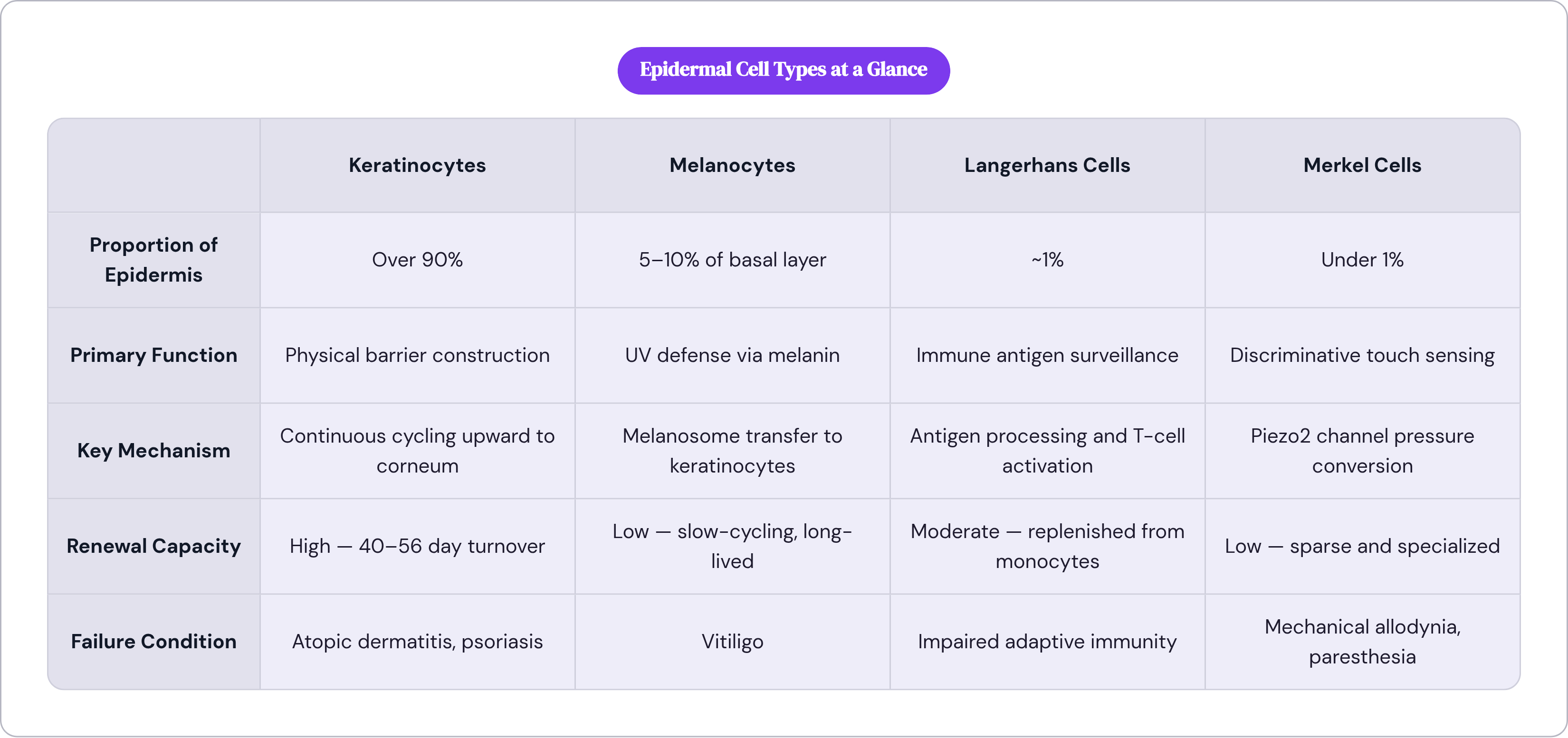
No single cell in the skin works alone. Keratinocytes signal Langerhans cells when threats appear. Melanocytes transfer melanin to keratinocytes so UV defense is distributed rather than concentrated. Merkel cells work in tandem with afferent neurons to complete the sensory circuit. Fibroblasts maintain the extracellular matrix that supports all the dermal populations embedded within it. Every functional capability in the skin is a collaboration.
The epidermis alone requires at least four distinct cell types to cover its core functions: barrier construction, UV defense, immune surveillance, discriminative touch. Those functions cannot be condensed into fewer cell types without losing capability. The cellular diversity isn't excess. It is the mechanism.
There's a distinction worth making between redundancy at the system level and specialization at the cell level. The skin has redundant immune capacity: Langerhans cells in the epidermis, mast cells, macrophages, and lymphocytes in the dermis, overlapping defensive coverage across layers. But within that system, each cell type is strictly specialized. Keratinocytes don't produce melanin. Melanocytes don't synthesize collagen. Merkel cells don't coordinate immune response. The robustness of the organ as a whole comes from distributing specific functions across specific cells, not from any cell's capacity to cover multiple jobs.
This changes how I think about what I see, clinically and otherwise. Slower turnover in aging skin isn't a surface texture issue. It's keratinocyte biology on a longer timeline. Loss of elasticity isn't a cosmetic abstraction. It's fibroblast output declining over years, collagen fragmenting, structure degrading from within. Depigmentation isn't a pigment deficit. It's a slow-cycling cell type destroyed without adequate replacement capacity.
Any framework for skin health, whether it's a topical product, a therapeutic intervention, or a diagnostic approach, that treats the skin as a uniform substrate will hit the ceiling of that assumption. The skin is specific by design, layer by layer, cell by cell, each one doing exactly what the others cannot.