Skin Layer Anatomy and Function
Your skin rebuilds its protective barrier completely every month, cell by cell.
How the epidermis builds its barrier one cell generation at a time
Start at the surface, because that is where everything is most exposed. The epidermis is the thinnest of the three layers and the one absorbing the most direct punishment: pathogens, UV radiation, chemical insult, mechanical abrasion. What makes it particularly interesting is that it handles this not through passive toughness but through a wall that tears itself down and rebuilds from the inside, continuously, without stopping.
About 95 percent of epidermal cells are keratinocytes, and what a keratinocyte does across its entire lifespan is a single, directional journey outward. It starts in the deepest sublayer, the stratum basale, through mitotic division. Then it migrates upward through the stratum spinosum, the stratum granulosum, and finally the stratum corneum. Along the way it flattens, sheds its nucleus, and loads up on keratin and lipids until it is, functionally, structural scaffolding. By the time it reaches the surface, it is ready to be shed.
The stratum corneum is 25 to 30 layers of these anucleate, keratin-dense cells. Your body sheds roughly 40,000 of them per day, and the mitotic activity back in the stratum basale replaces them on a cycle of approximately 28 to 30 days in younger adults, a timeline that stretches noticeably as we age. That means the barrier you are relying on right now is not the one you had a month ago. For something so central to how your skin works, that fact gets surprisingly little attention.
In the stratum spinosum, cells are held together by structures called desmosomes, which give the layer a spiny appearance under a microscope. That visual quirk is actually a mechanical solution: desmosomes keep cells tightly coupled under physical stress so the layer resists deformation rather than shearing apart.
At the stratum granulosum, cells release lipid-rich granules into the intercellular spaces. This is where the skin's waterproofing gets actively built. The moisture barrier marketed across every skincare aisle is not some passive property of the outermost surface. It is constructed one cell-generation deep, through deliberate secretory activity, before those cells even arrive at the stratum corneum.
The palms and soles have a fifth sublayer, the stratum lucidum, sitting between the granulosum and corneum. The regions bearing the most mechanical load get additional lamination. You see that logic repeat at every level of this anatomy: wherever demand is highest, the structure is more.
The specialized cells that give the epidermis functions beyond physical barrier
Keratinocytes build the wall. But three other cell types have established themselves within that wall to give the epidermis capabilities a wall alone cannot provide, and their exact positioning is not incidental.
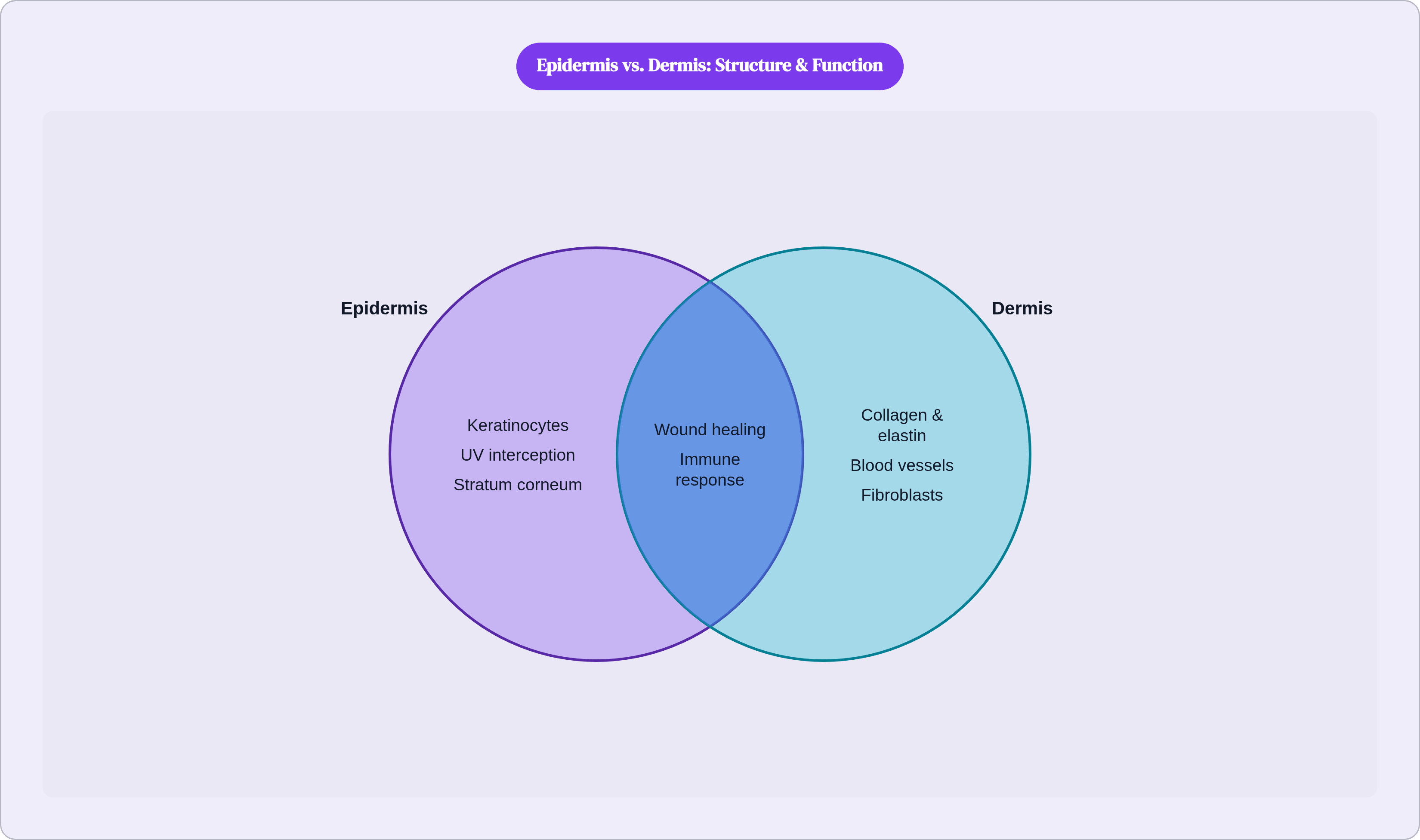
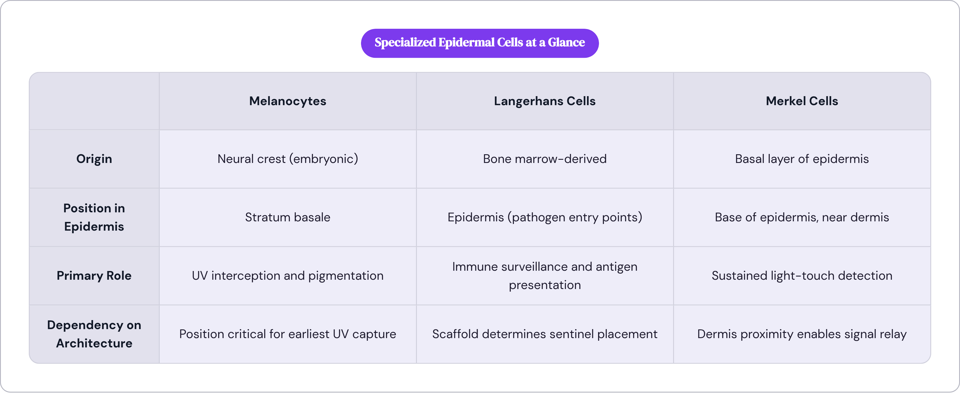
Melanocytes originate in the neural crest during embryonic development and settle into the stratum basale. They produce the pigment that gives skin its color. But their operationally significant role is intercepting UV radiation before it reaches deeper structures, and they do this from precisely the right position: at the base of the outermost layer, where they encounter radiation at the earliest possible opportunity.
Langerhans cells are immune sentinels, antigen-presenting cells that sample the environment, identify foreign proteins, and initiate immune responses. Their position in the epidermis places them exactly where pathogens are most likely to attempt entry. If the stratum corneum is breached, Langerhans cells are the next contact. They are bone marrow-derived and replenishable, but they depend entirely on the keratinocyte scaffold for their positioning. Disrupt the scaffold and you displace the sentinel.
Merkel cells are pressure sensors, concentrated at fingertips and lips where fine tactile discrimination matters most. They are slow-adapting, meaning they respond to sustained light touch rather than rapid change, and they sit near the base of the epidermis, adjacent to the dermis's nerve supply. They are stationed at the interface between the layer that contacts the world and the layer equipped to relay signals inward. The placement is doing real work.
None of these three cell types simply coexist with keratinocytes. Their function depends on epidermal architecture. Significantly disrupt the epidermis and you lose the physical barrier, yes, but you also displace cells whose effectiveness is entirely contingent on being in the correct stratum at the correct depth. That is two losses for the price of one.
Research published in the Journal of Translational Medicine in 2024 highlighted the role of epidermal stem cells at the basal layer in wound healing and immune-related conditions. The same mitotic machinery that replaces shed cells daily is also the machinery that responds to injury. One system, multiple outputs, all running through the same cellular infrastructure.
What the dermis contributes that the epidermis cannot supply itself
The epidermis has no blood supply. None. Every nutrient and oxygen molecule it uses has to diffuse upward from the dermis below. When I first really sat with that fact, it reframed everything: the dermis is not simply the middle layer. It is the epidermis's life support, and the epidermis is entirely dependent on the dermis in a way that only becomes visible when something disrupts that supply.
The dermis is the thickest of the three layers, dense with functional infrastructure: collagen fibers, elastin fibers, blood vessels, lymphatics, nerves, sweat glands, sebaceous glands, hair follicles. Two sub-regions define it. The papillary dermis sits at the top in loose connective tissue that interfaces directly with the epidermal basal layer. The reticular dermis sits deeper, with denser, net-like arrangements of collagen and elastin that give skin its tensile strength, its capacity to stretch and return.
Collagen and elastin are doing different mechanical jobs. Collagen resists tearing; elastin allows deformation and recoil. What most people do not realize about elastin is that it has a notably slow turnover rate, which means it accumulates damage across years rather than quietly repairing itself. Chronic UV exposure and simple aging degrade elastin fibers your body cannot rapidly replace. The visible consequence of that structural fact is something you may recognize in the mirror without connecting it back to a specific tissue mechanism.
Glycosaminoglycans, often abbreviated as GAGs, are water-binding molecules distributed through the dermis's extracellular matrix. They represent a small fraction of total skin weight, but their water-binding capacity is what maintains dermal volume and underlies visible plumpness. When GAG content decreases with age, the volume loss is direct, not metaphorical: it is a structural consequence of reduced water retention in the matrix.
Fibroblasts produce and maintain all of this extracellular matrix. They synthesize collagen, elastin, and GAGs; they repair the dermis after injury; and their activity declines with age in ways that have measurable downstream effects on skin structure. A fibroblast is not a passive cell in a stable material. It is an active maintenance worker in tissue that requires constant upkeep, and it slows down whether you want it to or not.
The dermis's blood vessels do double duty: they supply the epidermis above, which has no blood vessels of its own, through diffusion and connect downward to the larger vascular network of the hypodermis below. The dermis is literally the conduit between layers, the middle link in a supply chain that begins in the body's core and ends at the skin's surface.
How the dermis regulates temperature and communicates sensation
Thermoregulation is a whole-body problem. The dermis contributes two mechanisms, and they work differently enough that conflating them actually muddies how you think about skin's role in thermal management.
Eccrine sweat glands secrete water and sodium chloride onto the skin surface; evaporation of that secretion draws heat away from the body. Separately, dermal blood vessels dilate to increase heat loss through the surface or constrict to conserve it. Both are controlled responses to thermal load, not passive side effects of the dermis's structure. Your body is actively managing heat through a layer of tissue that is simultaneously doing a dozen other things.
Apocrine glands, concentrated in the axillary and genital regions, produce a different secretion, one metabolized by the skin's resident microbiota. The odor associated with these glands is a byproduct of microbial activity on that secretion, not the secretion itself. This is easy to get backwards. The skin is not just a physical structure; it is an ecosystem, and its surface chemistry emerges from the interaction between human biology and microbial community.
The sensory receptors distributed through the dermis are organized by depth, and that organization is doing real mechanical work. Meissner's corpuscles sit near the surface in glabrous (hairless) skin, particularly at the fingertips, and account for roughly 40 percent of the sensory innervation of the human hand. They are tuned to low-frequency vibration and fine texture: reading surfaces, distinguishing materials, manipulating small objects.
Pacinian corpuscles sit much deeper, extending into the hypodermis. Their architecture, concentric tissue layers surrounding a nerve ending, filters out static pressure and responds selectively to high-frequency vibration. The layered structure is not ornamental; it is a mechanical filter, and its depth in the tissue corresponds directly to the type of signal it processes. Ruffini endings detect skin stretch and joint deformation. Free nerve endings, the most abundant receptor type, extend as high as the mid-epidermis and handle pain and temperature.
Shallow receptors handle fine surface information. Deep receptors handle signals that require isolation from surface noise. The geometry of sensation is built into the anatomy, not added afterward.
What the hypodermis does that the layers above depend on
The hypodermis sits below the dermis and is sometimes excluded from formal definitions of skin. That classification debate is, frankly, less interesting than the functional reality: the boundary between dermis and hypodermis is not sharply defined anatomically, and the hypodermis performs functions the upper layers depend on in ways that only become obvious when something goes wrong.
Its primary tissue is adipose. Adipocytes cluster in groups held in place by connective tissue, forming a layer of mechanical cushioning between the skin and the underlying muscle and bone. This absorbs physical impact before it reaches deeper structures. The fat is not incidental padding. It is a shock-absorption system, and its absence, through disease, atrophy, or surgical disruption, makes that function immediately legible in ways that are hard to miss.
Thermal insulation is the second role. Fat conducts heat poorly, which means the hypodermis slows the rate at which body heat escapes to the environment. Hypodermal thickness varies with climate adaptation, body region, and individual physiology for exactly this reason. The variation reflects real thermoregulatory demands.
The connective tissue in the hypodermis anchors the dermis to the underlying fascia, muscles, and bones. Without it, the skin above would not stay in place during movement. Structural contributions that work seamlessly tend to be the ones we ignore until they fail. Disruption through surgery, injury, or disease makes the dependency visible very quickly.
Blood vessels and nerves from the dermis enlarge and branch in the hypodermis, connecting upward to the skin and downward to the systemic circulation. Fat distribution varies by body region, sex, and age, which is why hypodermal changes, such as atrophy with aging or lipodystrophy (abnormal fat loss or redistribution), visibly alter the appearance and behavior of the layers above in ways that no amount of surface treatment can fully reach.
How the three layers fail and recover together
The clearest way to understand how these layers relate is to watch what happens when they are stressed.
Consider a burn severe enough to destroy the epidermis. The immediate loss is the physical barrier, but the consequences do not arrive in sequence. Without the epidermis, dermal structures are exposed to pathogens. Fluid regulation fails. Thermoregulation is compromised because the evaporative surface is gone and the underlying vasculature is now exposed to the environment. These effects emerge simultaneously because the epidermis was simultaneously serving all of those functions. Lose one layer and you lose the outputs of the whole system at once, not one by one.
Wound healing follows the same logic, running in the other direction. Epidermal stem cells at the basal layer resurface the wound. Fibroblasts in the dermis rebuild the extracellular matrix. Hypodermal vasculature supplies the blood flow that makes both possible. The surface cannot heal without support from below; rebuilding the deeper matrix requires the surface protection above. Recovery is coordinated across layers because the original structure was coordinated across layers.
Chronic sun exposure offers a more gradual illustration. UV radiation damages melanocytes in the epidermis, compromising pigment-based protection. Separately, it degrades elastin in the dermis, a tissue with slow turnover that accumulates damage across years rather than repairing discreetly. Neither effect alone explains why photoaged skin looks and behaves as it does. The combination, compromised protection at the surface and structural degradation beneath it, produces results qualitatively different from either insult in isolation. Treating photoaging by addressing only one of those layers will tend to fall short of addressing the other.
Inflammatory conditions like eczema and psoriasis often originate in disrupted epidermal barrier function, but they involve dermal immune activity and vascular changes as well. Treating the surface addresses part of the problem while the underlying dermal involvement continues. This is why these conditions persist the way they do: a surface-level intervention applied to a multi-layer problem will be incomplete, not because the intervention is wrong but because it is not aimed at enough of the system.
The skin microbiome underscores all of this. Microbial disruption at the epidermal surface triggers immune cascades that reach the dermis and implicate the vascular and lymphatic systems embedded there. An event that begins at the outermost surface propagates inward, across layer boundaries that textbooks draw as clean lines and the body never actually recognizes as such. That mismatch between how we diagram skin and how skin actually behaves is, I think, the most practically important thing to carry away from all of this.