How Skin Barrier Function Works
The lipid ratio in your skin barrier matters more than the individual ingredients.

How the stratum corneum builds a physical seal from dead cells and fat
You've probably encountered the brick-and-mortar analogy. It's accurate, and it repays more careful attention than it usually gets.
The bricks are corneocytes: dead, anucleate cells that were once living keratinocytes. Over the course of differentiation, they shed their nuclei and most of their organelles, packed themselves with structural proteins, and flattened into dense, overlapping plates. Mechanical integrity, some UV mitigation, hydration regulation. Purpose-built in a way that still strikes me as elegant, even after years of looking at this stuff.
The mortar is where it gets interesting. Not generic fat. A specific mixture of ceramides, cholesterol, and free fatty acids, arranged in lamellar bilayer structures, the kind of precise molecular architecture that physically prevents water from diffusing outward and foreign agents from penetrating inward. The ratio of these three lipids matters as much as their individual presence. When the ratio drifts, the bilayer structure changes, and barrier function changes with it. That is not a subtle distinction. It is the practical difference between a product that works and one that fails, even when both claim ceramides on the label.
A 2024 finding in the International Journal of Cosmetic Science made this visible: changes in ceramide level and composition appear consistently across multiple skin conditions. The lipid layer is not inert. It shifts in response to disease, stress, environment. Dynamic, in other words, which means vulnerable in ways most people rarely consider when they reach for a moisturizer.
A 2025 molecular dynamics study in Molecular Pharmaceutics pushed this further. Not all ceramides behave the same way. Ceramide NS is associated with impaired barrier function; ceramide NP with a healthy one. So when a product claims to contain ceramides, the relevant follow-up questions are: which ones, in what ratio, and doing what in the bilayer. Most labels will leave those questions unanswered.
What filaggrin does and why its absence destabilizes the whole structure
Filaggrin earns the word keystone because it does two structurally distinct jobs, and both are load-bearing.
First, it aggregates keratin filaments within corneocytes, giving them structural coherence. Second, it breaks down into the components of natural moisturizing factor, the hygroscopic molecules that keep the stratum corneum hydrated from within. When filaggrin is absent or reduced, both jobs fail simultaneously, and that simultaneity is what makes its loss so destabilizing. It is not one domino falling; it is two separate pillars giving way at the same moment.
The consequences cascade from there. Keratin filaments become disorganized. Lamellar body loading is impaired, meaning the lipid ratio in the mortar gets disrupted before cells even finish differentiating. Lamellar bilayer architecture turns abnormal. Corneodesmosome density drops, weakening cell-to-cell adhesion. And because filaggrin breakdown contributes to the skin's acidity, its absence raises stratum corneum pH.
That pH shift is not cosmetic. Healthy skin surface acidity, roughly 4.5 to 6.0, activates the enzymes that process ceramides and regulates barrier activity broadly. When pH rises, those enzymes underperform, lipid processing degrades further, and pro-inflammatory mediators are more readily released by keratinocytes. A single genetic variant destabilizing lipid organization, pH regulation, and inflammatory signaling simultaneously is part of why filaggrin loss-of-function mutations are among the strongest known genetic risk factors for atopic dermatitis.
One clarification worth making here: filaggrin deficiency does not directly break tight junctions. It enables the inflammation that does. That sequence changes how you think about disease progression, about where in the timeline intervention makes the most sense.
Tight junctions: the living checkpoint beneath the dead-cell layer
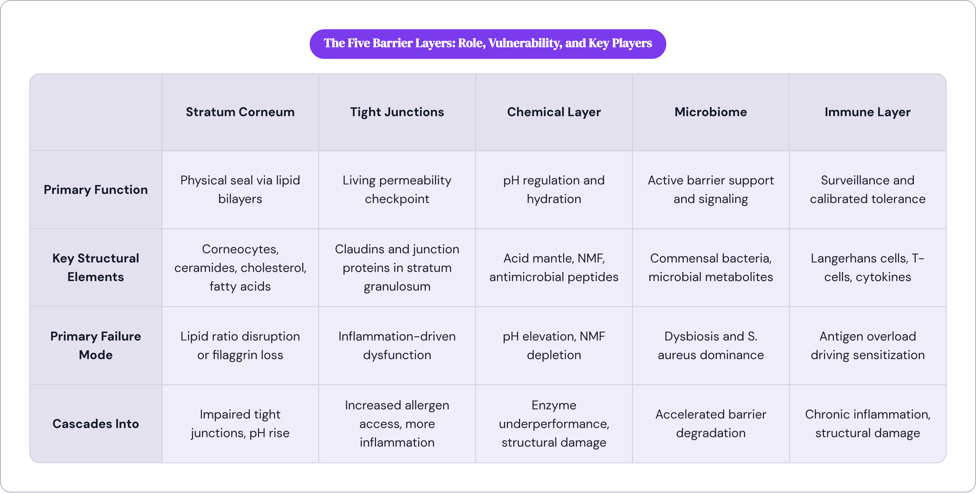
Tight junctions sit in the stratum granulosum, the outermost living layer, just beneath the stratum corneum. Unlike corneocytes, they are not structural in a static sense. They respond. They modulate permeability in real time, which is what makes them a genuine barrier layer rather than passive scaffolding.
Here is where the interdependence stops looking like a neat sequence and starts looking like something messier. Inflammation generated by filaggrin deficiency induces tight junction dysfunction. Tight junction dysfunction further impairs the stratum corneum above it. Greater permeability means more allergen exposure. More allergen exposure drives more inflammation, which further disrupts tight junctions.
There is no moment where this self-corrects without intervention. The research literature is fairly unambiguous on that point, even if it is not always framed so bluntly. Structural defects in tight junctions do not stay confined to the physical layer either; they feed directly into immunological impairment, which is where the next set of complications begins.
The acid mantle, NMF, and antimicrobial peptides as the skin's chemical defense layer
The chemical layer gets collapsed into "moisturizer" in most consumer conversations, which strips out most of what it actually does.
Start with pH. Healthy skin surface sits between roughly 4.5 and 6.0, an environment hostile to many pathogens and essential for regulating the ceramide-processing enzymes mentioned earlier. This acidity is not a byproduct. It is functionally necessary. When pH rises, whether from filaggrin loss or from a harsh alkaline cleanser, the chemical layer loses its regulatory capacity before any structural damage is even visible. That timing matters if you are trying to understand why some people's skin reacts to products that appear, on paper, perfectly gentle.
Natural moisturizing factor is a collection of hygroscopic molecules, ones that attract and hold water, inside corneocytes: amino acids, lactic acid, urea, pyrrolidone carboxylic acid, among others. They draw and retain moisture from within the cell. When barrier function is compromised, NMF levels fall, and the skin loses this internal moisture-drawing capacity, compounding the dehydration already caused by lipid disorganization. Two mechanisms pushing in the same direction at the same time.
Antimicrobial peptides, cathelicidin LL-37 and human beta-defensins hBD-1, hBD-3, and S100A7 among them, provide broad-spectrum activity against bacteria, viruses, fungi, and some parasites. But they also upregulate tight junction proteins, directly reinforcing the physical layer. That second role gets overlooked in most discussions. The chemical and physical barriers are not exchanging occasional signals across a gap; they are the same system operating at different organizational levels. Sebum-derived lipids contribute here as well, though the full accounting of their role is still being worked out.

The skin microbiome as an active barrier participant, not a passive resident
The microbiome establishes itself immediately after birth and is constitutive to how the immune system learns to function. It is not a later addition to the barrier. It is present from the beginning, woven into the architecture before the architecture is even fully formed.
Staphylococcus epidermidis, a common commensal, increases skin ceramide levels and reduces transepidermal water loss through a sphingomyelinase-dependent mechanism, at least in mouse models. A bacterium living on the skin surface is directly supporting the lipid component of the physical barrier. The microbiome and the stratum corneum are in active, ongoing communication, not merely coexisting.
During atopic dermatitis flares, microbial diversity collapses. S. aureus dominates, producing proteases and toxins that degrade an already compromised barrier. Dysbiosis is not merely a consequence of barrier dysfunction, sitting passively alongside it. It accelerates the dysfunction. Microbial metabolites, short-chain fatty acids and indole derivatives among them, influence cutaneous immune responses, lipid metabolism, and barrier integrity through metabolite signaling. The microbiome has a chemical vocabulary it uses to communicate with both the immune and chemical layers simultaneously.
It is also the most environmentally sensitive layer. Antibiotics, detergents, humidity, geography: all shift microbial populations faster and more dramatically than they shift filaggrin expression. That sensitivity makes the microbiome both the most tractable element of the system and, in some ways, the most fragile. It's the first thing to shift when something goes wrong, and harder to stabilize once it has shifted than you might expect.
How the immune layer monitors, tolerates, and responds without overcorrecting
The epidermis contains Langerhans cells, tissue-resident memory T-cells, and gamma-delta T-cells. Their collective job is immune surveillance: sampling what passes through the barrier, distinguishing harmless antigens from genuine threats, and maintaining that distinction consistently across years of cumulative exposure.
"Tolerance" is underused in popular discussions of skin immunity. The immune layer's function is not only defense. It is calibrated tolerance, deciding what to ignore as much as what to engage. That calibration depends heavily on the integrity of the layers above it. When the physical and chemical barriers are intact, relatively few antigens reach immune cells, and measured, appropriate responses are the likely outcome.
When those layers are compromised, more antigens arrive, and the immune layer was not designed for that volume. Sensitization becomes more probable. Inflammatory responses that serve no protective purpose, and that cause direct structural damage, become more frequent. The system overreacts because it is receiving a signal it was not built to process at that scale, not because something is fundamentally wrong with the immune layer itself.
The research literature uses "extensive crosstalk" to describe how the immune layer operates, and that framing resists the instinct to locate the problem somewhere specific. The immune layer receives signals from every other layer and generates new signals that affect every other layer. It is simultaneously sensor, effector, and signal source. This is part of why inflammatory skin diseases like atopic dermatitis and psoriasis are so resistant to single-target interventions. The problem is not localized. It is distributed across the architecture, and treating it as though it has a single address is how you end up with therapies that work briefly and then stop.
How TEWL measurements make barrier function visible and what they actually capture
Transepidermal water loss, TEWL, measures water vapor diffusing across a fixed area of stratum corneum per unit time. It is the most widely used objective measurement for assessing physical barrier function, and it is a direct readout, not a proxy.
Atopic dermatitis patients show measurably higher TEWL and lower stratum corneum hydration, quantifiable confirmation that the structural disruptions described in earlier sections have observable, detectable consequences. It also means you can track change over time, which is more useful than before-and-after photographs, and more honest.
TEWL has real limitations. It is sensitive to body site, ambient temperature and humidity, recent physical activity, and time of day, all of which make standardizing conditions difficult and make cross-study comparisons require careful scrutiny. Electrical impedance spectroscopy is an emerging alternative that measures permeability through electrical response across multiple frequencies, offering better precision in variable conditions, though it has not yet displaced TEWL as the standard.
The reason this matters for you is not primarily technical. Barrier function is not theoretical. It can be measured and tracked. That means the question of whether a repair strategy is actually working, rather than just producing cosmetically appealing photographs, is answerable. And the answer, increasingly, is being established rather than assumed.
What actually damages the barrier, and why the damage tends to compound

Damage enters the system from multiple directions simultaneously. Genetic variants, environmental stressors, psychological stress, nutritional insufficiency, and pollutants each engage the barrier at different points and through different mechanisms, which is one reason single-cause explanations for conditions like eczema have been incomplete.
Environmental pollutants make this concrete. When PM2.5 particulate matter is applied to a human three-dimensional skin model, expression of keratin 10, desmocollin 1, and claudin 1 decreases. These are structural barrier proteins. Airborne particulate matter is not just accumulating on the skin surface; it is downregulating the molecular architecture of the barrier at the gene expression level, before you've touched your skin, before you've changed anything about your product routine.
Psychological stress connects to barrier function through neuroimmune pathways. The neuronal layer receives the least attention in consumer-facing skin education, partly because the mechanisms are still being mapped. Mental state and skin permeability are not metaphorically linked. They are mechanistically connected, and the full picture of those connections remains incomplete. That's not a comfortable place to sit if you want clean answers, but it's where the science currently is.
The compounding pattern runs roughly like this: filaggrin deficit raises pH; elevated pH disrupts ceramide-processing enzymes; lipid disorganization weakens the stratum corneum; a weakened stratum corneum invites microbial imbalance; dysbiosis triggers inflammation; inflammation breaks tight junctions; impaired tight junctions increase antigen access; greater antigen exposure worsens immune dysregulation; immune dysregulation drives more inflammation, which loops back to lipid organization.
It is a loop, not a chain. That structural difference explains why barrier diseases tend toward chronicity rather than acute resolution. Interrupt the loop at one point and the pressure does not dissipate. It continues from every other point simultaneously.
How barrier repair works with the layer structure rather than targeting a single component
Repair logic follows the architecture. Multi-component by necessity, not because more ingredients make a better product, but because the system being repaired has multiple independent failure points that do not wait for each other to resolve.
The ceramide-to-cholesterol-to-fatty acid ratio recommended by dermatological societies for optimal barrier restoration is 3:1:1. The specificity of that ratio reflects what molecular research on lamellar bilayer formation has established: composition determines function. Not presence, not quantity alone, composition. A product can contain all three lipids and still underperform if the ratio is wrong.
A 2022 randomized controlled trial in the British Journal of Dermatology found that enhanced lipid chain ordering was significantly associated with barrier integrity, with a correlation coefficient of 0.61. Treatment with the test cream increased hydration by 8.61 capacitance units and reduced dryness signs compared to the reference. That granularity illustrates what rigorous evaluation of "barrier repair" actually looks like: the outcome is measurable, the mechanism is traceable, and neither has to be taken on faith.
Antimicrobial peptides like LL-37 and human beta-defensins improve tight junction barrier function directly, which means supporting the chemical layer has structural consequences for the physical layer above it. These are not independent levers. They are connected outputs of the same system.
The microbiome's role in repair remains an active research frontier. How to support commensal populations in ways that meaningfully contribute to barrier restoration is not settled, and distinguishing legitimate inquiry from the marketing claims that have already rushed into that space takes some care and a reasonable tolerance for ambiguity.
What the layered model ultimately clarifies is why single-ingredient interventions so often disappoint and then get abandoned. The barrier being repaired is a system with multiple interdependent failure points. Addressing one while the others continue cycling is not a treatment strategy. Consistent, multi-component support is not a compromise or a hedge. It is what the architecture actually requires.


