What AI Skin Tools Can and Cannot Diagnose
These tools excel at common skin cancers but fail on rare conditions and darker skin tones.
The access problem is real before we even talk about the technology. The United States has roughly 3.4 dermatologists per 100,000 people. That ratio looks worse the moment you leave a major city. Around 5 million skin cancers are diagnosed in the U.S. every year, and early detection meaningfully changes outcomes for most of them. The teledermatology market hit roughly $15.76 billion in 2024 and is projected to approach $63 billion by 2032. That is not hype money; that is demand chasing a real shortage. COVID-19 accelerated the shift toward remote care, and that comfort with digital health tools did not reverse when clinics reopened. Millions of people now photograph spots, rashes, and lesions and submit them to apps before or instead of seeing a doctor.
So the question is not whether there is a use case. There clearly is. The question is how well these tools actually perform, where they break down, and what it costs when they do.
How AI Skin Tools Actually Work
Most consumer-facing skin tools run on convolutional neural networks, or CNNs, trained on large labeled image datasets. The model learns to associate visual patterns in images with diagnostic labels. Simple in concept, surprisingly powerful in practice when the conditions are right.
Some clinical devices go further than photography. DermaSensor, FDA-cleared in January 2024, analyzes light scattered back from a skin lesion. Cancerous and benign tissue give off different light signatures because cellular changes alter how tissue interacts with light at the molecular level. That is a different class of tool than a smartphone app with a camera.
Large language models, meaning ChatGPT, Claude, and similar tools, work through a different mechanism entirely: text and image reasoning rather than dedicated visual classification. They are worth distinguishing from purpose-built dermatology models, because they are generalist tools being applied to a specialist problem, with predictable consequences.
The constraint baked into every one of these systems from the beginning is this: they learn from images and labels. Their knowledge is only as broad and representative as the datasets they were trained on. That single sentence explains most of what follows.
Output from these tools is typically a probability score or risk classification, not a clinical diagnosis. That distinction matters legally, clinically, and practically.
What AI Does Well: Pattern Recognition in Skin Cancer and Common Inflammatory Conditions
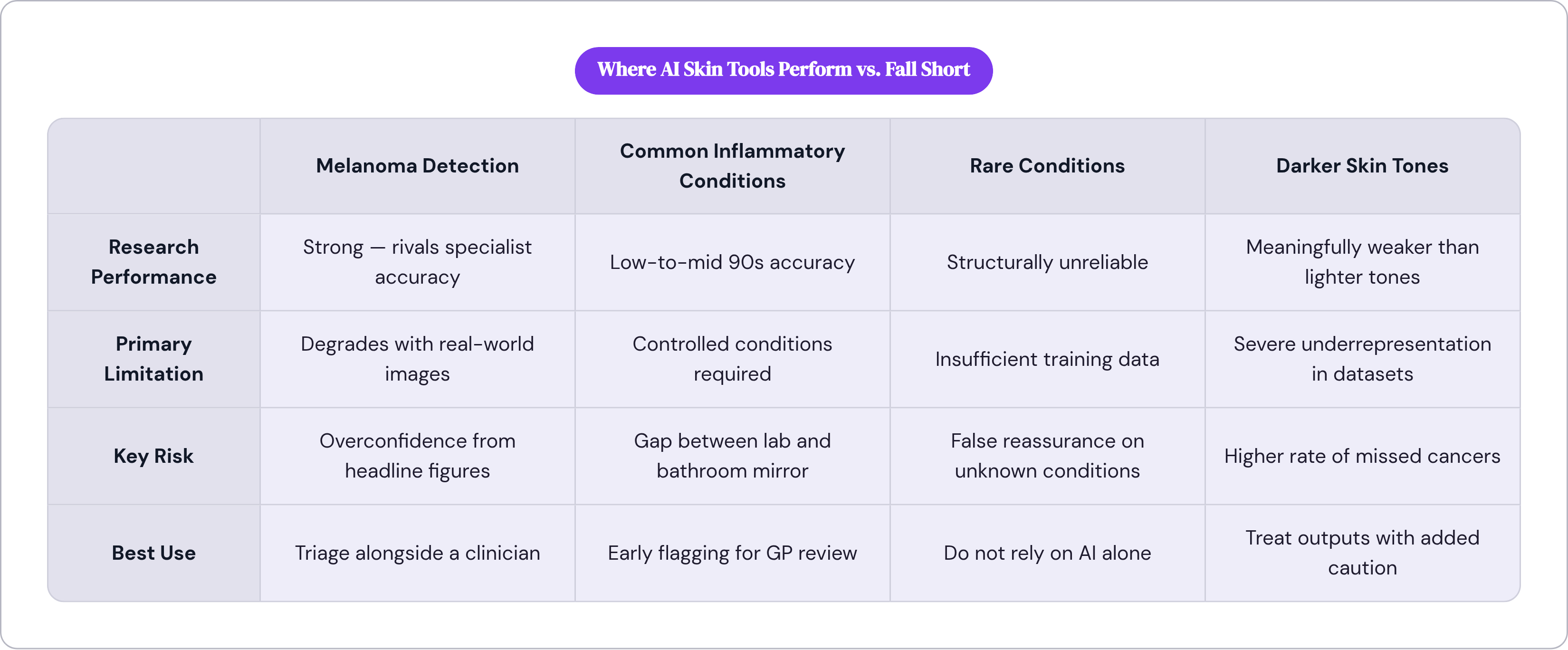
The numbers for melanoma detection, under ideal conditions, are striking. A 2025 review covering 551 studies found CNNs achieved 91% sensitivity and 94% specificity distinguishing melanoma from benign lesions. For context, a large 2024 meta-analysis found dermatologists using dermoscopy (a magnified skin examination tool) achieved 85.7% sensitivity and 81.3% specificity. AI's best results are now statistically comparable to specialist performance. That is not marketing copy; that is the peer-reviewed literature.
Common inflammatory conditions also show strong performance in research settings: accuracy in the low-to-mid 90s for acne and rosacea, similar figures for eczema, and high 80s for psoriasis.
DermaSensor, in a Mayo Clinic-led trial, demonstrated 96% overall sensitivity for malignancy, compared to 83% for primary care physicians. Its sensitivity specifically for melanoma came in at 95.5%.
Real-world triage volume matters here too. In May 2024, one AI triage platform analyzed over 18,000 cases and flagged a meaningful number of melanomas, squamous cell carcinomas, and basal cell carcinomas, amounting to roughly 5% of the UK's monthly melanoma detections. At scale, that is a non-trivial contribution to early detection.
A 2024 meta-analysis found that when general practitioners used AI support, they achieved 90% sensitivity and 85% specificity for detecting suspicious pigmented lesions. That is the clearest articulation of the use case: AI as a triage layer in primary care, catching things that might otherwise wait months for a specialist.
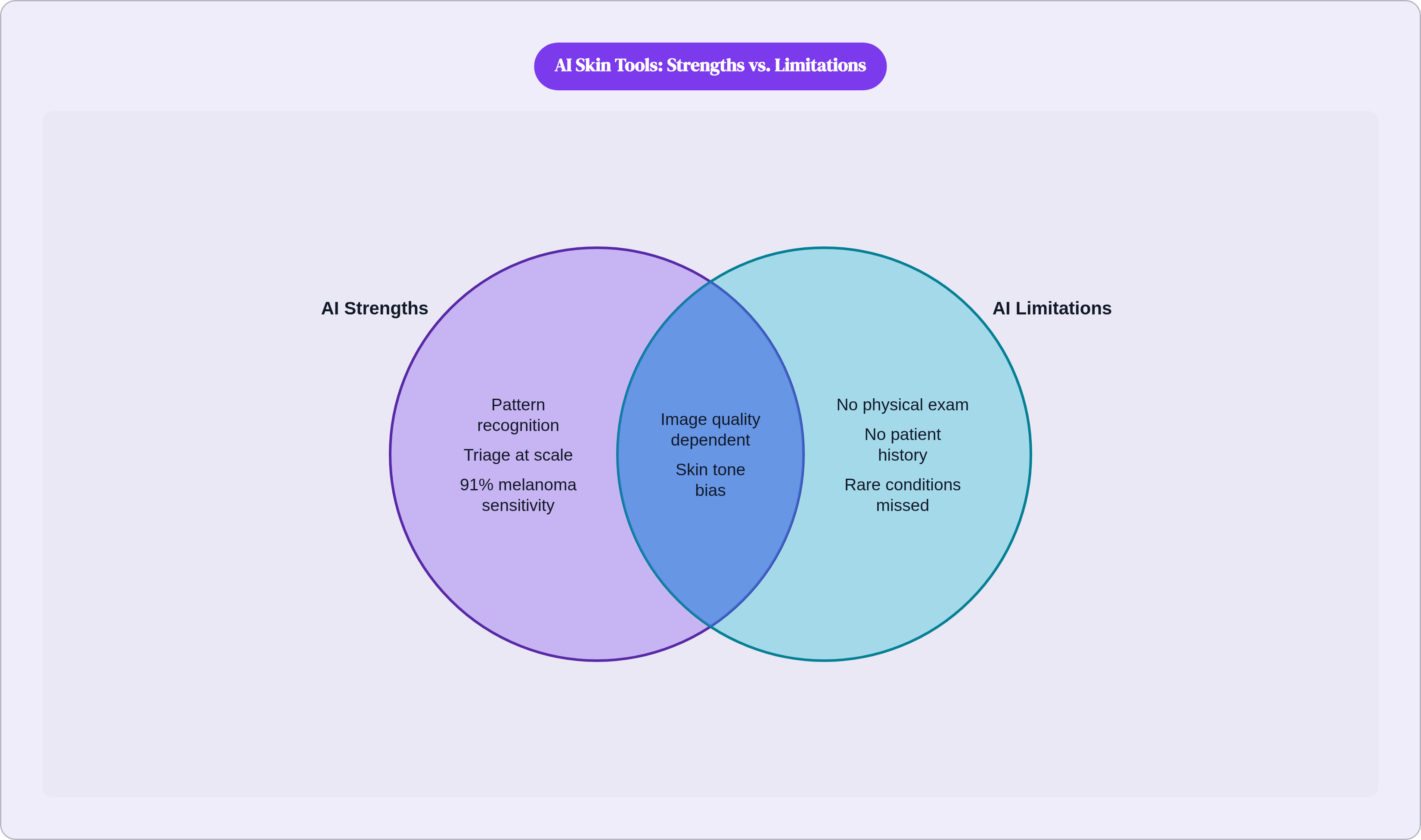
The strength is pattern recognition at scale across known, well-represented conditions. Hold that qualifier.
Where Performance Falls Apart: Image Quality, Rare Conditions, and Real-World Variability
Those headline accuracy figures come from controlled studies using high-quality, standardized images. Real-world smartphone photos introduce lighting variation, blur, shadow, scale ambiguity, and partial obstruction. The controlled setting and your bathroom mirror are not the same thing.
A 2024 systematic review of AI in primary care found sensitivity ranging from 58% to 96.1% and accuracy spanning an enormous range. That spread tells the real story: controlled conditions produce the high end; everyday use produces the low end.
Specific anatomical locations remain difficult. Hairy scalp, mucous membranes, and nail beds pose recognized recognition challenges for current models. If the lesion is in one of those locations, your confidence in AI output should drop accordingly.
Rare conditions are structurally underrepresented in training data. This is not fixable by making a better model; it is a data problem. If the model has seen few examples of a condition, it cannot reliably classify it.
LLMs have their own distinct failure modes. In documented testing, one major LLM incorrectly labeled some melanomas as benign; another frequently misclassified nevi as melanomas. Neither is reliable as a standalone diagnostic tool. A 2025 survey found that 97% of dermatologists who use AI chatbots report having to manually edit responses before use. When professionals who understand the domain are correcting the output that often, the practical reliability ceiling for lay users is worth considering carefully.
Binary confidence scores, "high risk" or "low risk," also frequently lack the granularity that nuanced decisions require. A clinician doesn't just need to know risk level; they need to understand what pattern the model is responding to and how much confidence to assign given the image quality and context.
The Skin Tone Gap: How Training Data Bias Produces Unequal Performance
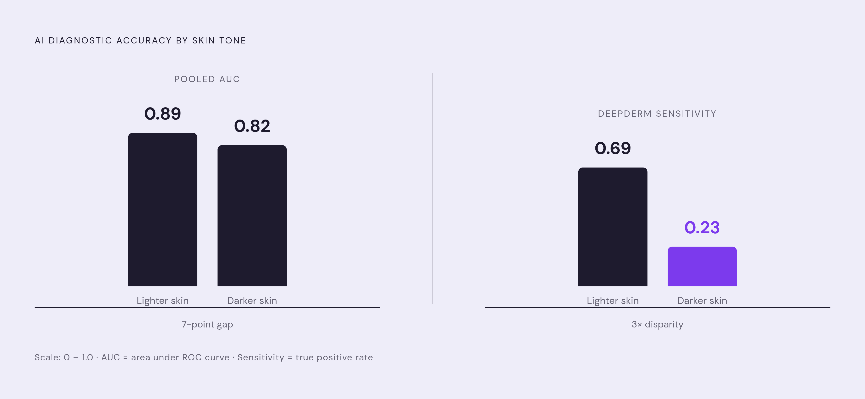
This is where the technology's limitations become a justice issue, not just a technical one.
A 2025 meta-analysis found AI systems achieved a pooled area under the receiver operating curve of 0.89 for lighter skin tones and 0.82 for darker skin tones, a seven-point gap that translates directly into missed diagnoses. Stanford's DeepDerm showed sensitivity of 0.69 for lighter skin tones and 0.23 for darker; nearly a three-fold disparity. ModelDerm showed similar drops.
The source of this disparity is in the data. Among over 106,000 clinical images analyzed in one study, only 11 images represented darker skin, with no representation from African, African-Caribbean, or South Asian populations. The data problem is foundational, not incidental. Among thousands of AI-generated dermatology images studied in 2025, only about 10% reflected dark skin, and only 15% accurately depicted the intended condition. The model can only learn what it has been shown.
The pattern that makes this particularly dangerous: models are more likely to misclassify cancerous lesions as benign in darker-skinned individuals. The primary risk is underdiagnosis, not overdiagnosis. A tool that misses cancer in one population while catching it in another is not a neutral shortcoming; it is a stratified failure.
Here is what matters: when models were trained on more diverse datasets, accuracy on darker skin tones improved. The fix exists. It requires deliberate action, specifically in how training data is collected and curated.
An ethical analysis published in the British Journal of Dermatology found at least one patient-facing app that explicitly excluded darker-skinned individuals as a target user. That is not a technical limitation; that is a design choice, and a disclosure that should inform how any user evaluates the tool.
If you have a darker skin tone, current AI skin tools are demonstrably less reliable for you. That is not a caveat buried in fine print; it is the most important thing to know before you use one.
What AI Cannot Do Regardless of Image Quality or Skin Tone
Some limits are not about training data or model architecture. They are structural to what image-based AI is.
AI cannot examine a lesion physically. No texture, firmness, warmth, or border elevation information is available to a model processing a photograph. A dermatologist's hands and dermoscope access dimensions of a lesion that a camera simply cannot capture.
AI has no access to patient history unless explicitly entered, and even then, it cannot interrogate that history the way a clinician does. Medication exposure, autoimmune conditions, recent infections, pregnancy, immune suppression: all of these can make identical-looking rashes mean completely different things. A rash is a surface event that can be the output of dozens of different systemic processes.
No tool can perform or order a biopsy. Biopsy remains the only definitive confirmation for malignancy. An AI risk classification is not a substitute for a biopsy and lab analysis.
Systemic conditions that manifest on skin, including lupus, drug reactions, and vasculitis, require labs, full history, and clinical judgment. A skin image captures only the surface of what may be a systemic problem that requires workup the image cannot prompt.
These are not gaps that model improvement will close. They are inherent to what image-based tools can do.
How to Use AI Skin Tools in a Way That Matches Their Actual Strengths
The strongest use case is triage: using a tool to help you decide whether something warrants a clinical visit, especially when your access is limited or your wait is long. That is a legitimate and valuable function.
Know which type of tool you are using. DermaSensor-style devices deployed by a trained primary care physician represent a different and more reliable context than a consumer app used alone at home. The tool, the operator, and the setting all matter.
A low-risk result is not clearance. It means the pattern in that image, under those conditions, does not strongly suggest malignancy. If something is changing, itching, bleeding, or simply worrying you, see a clinician regardless of what a tool says.
If you have darker skin, treat AI outputs with additional caution given the documented performance gaps. Do not let an AI risk score replace your own concern or a physician's clinical evaluation.
Image quality affects what the tool can detect. Practical steps help: take photos in natural light, ensure the lesion is unobstructed, include a reference object for scale. These things directly influence accuracy.
DermaSensor, used by primary care physicians, reduced the rate of missed skin cancers from 18% to 9%. That is the right model: AI as a support layer for a clinician, not a replacement for one. The clearest evidence consistently points in the same direction. Bring AI outputs to a clinical conversation; do not use them to close the question.
What Would Make AI Skin Tools More Trustworthy Over Time
Training data diversity is the most direct lever. Improving representation of darker skin tones and underrepresented populations in datasets demonstrably improves performance for those groups. The barrier is not technical; it is organizational and ethical.
Regulatory oversight matters. The FDA cleared DermaSensor in January 2024 as the first AI device indicated for use by non-specialists. That is a meaningful benchmark; it required demonstrated clinical validity. Most consumer apps operate outside that oversight entirely, with no obligation to prove they work. That gap is significant.
Transparency about limitations is not a nice-to-have. Tools that communicate uncertainty honestly, including what population they were validated on, under what imaging conditions, and with what confidence intervals, are more useful than tools that project false precision. Binary risk classifications that obscure the conditions under which that risk was computed can lead to overconfidence in either direction.
Integration with care, rather than separation from it, is what the evidence supports. The tools that demonstrably improve outcomes are those used alongside clinicians, not as a substitute for them.
For you, right now, trustworthiness is a practical question, not an abstract one: Does this tool have regulatory backing? What population was it validated on? Are you using the output as one input into a clinical conversation, or as a verdict? The answers to those three questions determine how much weight the output should carry.