How Clinical Training Scale Separates Purpose-Built Dermatology AI From General Medical Image Tools
Broader training data and clinical diversity separate specialized dermatology AI from general tools.
Purpose-built dermatology AI is best judged by the volume, diversity, and clinical detail of its training data. Multimodal clinical AI trained on labeled dermatology cases can connect skin images with patient history and diagnoses, but reliable use depends on matching the conditions and populations it will encounter. Clinicians must still oversee care when those predictions guide it.
Why the training data behind a dermatology AI model determines its clinical usefulness
A patient submits a photograph of a lesion, and an AI tool flags it for further assessment. What makes that flag clinically useful? The model needs to have learned from conditions, skin types, and clinical contexts that match the patient seeking care. A result on a familiar test dataset cannot answer that question by itself.
Because so much of dermatology depends on what can be seen, it was an early adopter of AI image analysis, which showed both promise and failure. General medical image tools learn broadly across modalities, including radiology, pathology, and ophthalmology, while dermatology-native models concentrate on skin. Their annotations can include lesion morphology, patient history, Fitzpatrick skin type, and coded diagnoses. Global dermatologist density is extremely low, with a disparity of more than an order of magnitude between high-income and low-income nations, so tools proposed to fill the gap need more than convincing demonstrations.
How early CNN-based tools exposed the limits of shallow dermatology training
Early convolutional neural networks, or CNNs, showed that AI could match specialists on selected dermatology benchmarks. Performance did not reliably transfer across datasets, conditions, or clinical settings. Public dermatology datasets spanned a wide spectrum, from the very small to the tens of thousands. Consequently, two tools described as “trained on dermatology data” could have learned from very different slices of clinical practice.
Recognizing conditions represented in a benchmark was not the same as assessing the broader range a dermatologist encounters. Experienced dermatologists and residents also operate at different levels, making a claim of “dermatologist-level” performance incomplete without the task and comparator. A narrowly trained model could match one tier without generalizing across clinical work. The scope of what it had learned constrained its performance.
That history also separates predictive success from interpretability. A model could produce an accurate answer in an experiment without making its reasoning clinically clear. More representative training addresses the breadth of recognition, while clinically specific labels make the learned task more closely reflect medical distinctions. Neither achievement, by itself, proves that a tool can replace a dermatologist.
What AI tools are trained on large dermatology datasets, and why does scale help?
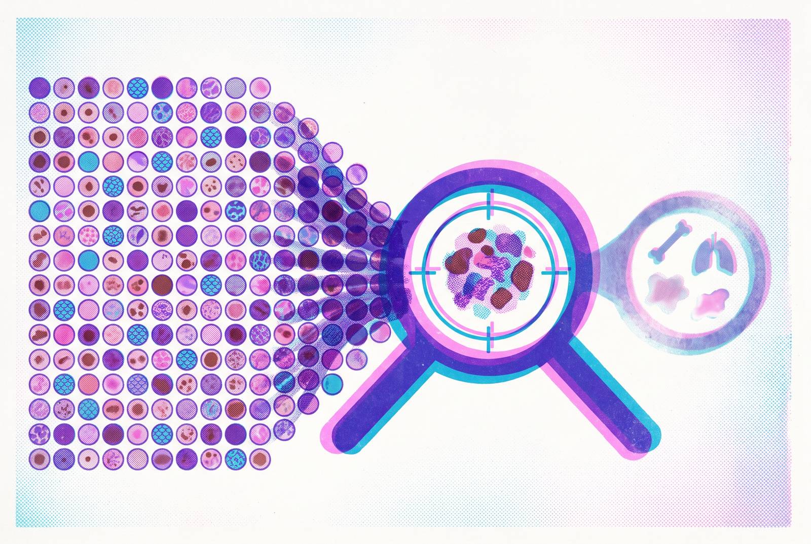
PanDerm and another model illustrate the move toward dermatology-native foundation models. DermFM-Zero remains a preprint, so claims about what it can do still need to be confirmed before they can count as settled clinical evidence. Models are now being trained on millions of real-world, multimodal dermatology images that come with clinical context. Scale alone is not the full explanation. The gain comes from combining volume with the range of skin presentations and the specificity of the information attached to them.
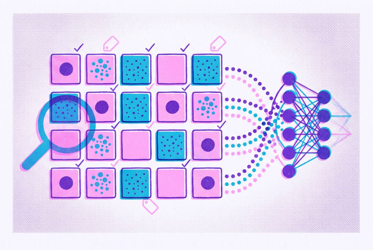
Clinical annotation supplies distinctions that raw photographs cannot provide on their own. Lesion morphology labels describe what can be seen, while patient history adds what the image alone cannot show. Fitzpatrick skin type classification and coded diagnoses add other clinically relevant structure. A large collection of photographs is therefore not interchangeable with a large collection of labeled clinical cases. Counts of images should also be kept separate from counts of patients or fully annotated cases.
The foundation-model approach has reported state-of-the-art performance with only a fraction of the labeled data required by prior approaches. This suggests that what a model learns from pretraining can help with later clinical tasks without having to learn recognition from a narrow dataset each time. It does not make labels unnecessary. The quality of the starting data can also shape what a model learns from later examples.
DermFlow provides a particularly revealing comparison with a general-purpose model. On biopsy-proven pigmented lesions from Indiana University clinics, it achieved substantially higher any-diagnosis accuracy and sensitivity than Claude Sonnet 4. However, the comparison has an important qualification. Claude received images only, while DermFlow also received patient histories, so the result does not isolate training specificity as the sole cause. It supports the value of dermatology-focused, context-rich assessment, but it cannot separate the effects of specialized training from the additional clinical information supplied at assessment.
Why generalization fails across real patient populations, and what the training data actually explains
If a model performs worse in a different population, what exactly changed? Skin tone is an important possibility, but so is the mix of diseases. A cancer-focused training collection may be poorly suited to a service where inflammatory and infectious conditions dominate. These are distinct shifts with different remedies. Adding more skin tones without broadening disease coverage can leave the central recognition gap intact.
Kunwor et al. 2026 separated these effects by evaluating a cancer-trained baseline, two open dermatology foundation models, and a general-purpose vision model across datasets stratified by skin tone and disease type. The cancer-specialized model lost most of its balanced accuracy on conditions it had not learned to recognize, with performance falling toward chance. Within the same diseases, skin-tone gaps were comparatively small and did not move consistently in one direction across models. The study used US-sourced datasets as controlled proxies for resource-constrained settings. What the study suggests for those settings must be inferred from its findings.
The failure also appeared before a classifier was trained. Cancer-specialized features barely grouped unfamiliar conditions, while dermatology-pretrained features retained substantially more transferable structure. This points to a representational gap. A model trained predominantly on neoplastic conditions may lack useful distinctions for eczema, tinea, impetigo, and scabies. Those conditions are central to primary-care and teledermatology work in resource-constrained environments.
Darker Fitzpatrick types are underrepresented in published dermatology AI research, and a documented deployment showed sharply reduced accuracy on deeply pigmented skin. That is a clinical safety gap affecting already-underserved patients. Disease coverage concerns whether the model knows the condition a patient has, while population coverage concerns how reliably it recognizes that condition in all the people seeking care.
What happens when purpose-built AI meets real clinical infrastructure: three documented deployments
Skin Analytics’ DERM system brings the autonomy question into clinical practice. A 16-month evaluation at two NHS hospital sites looked at autonomous triage of urgent suspected skin cancer referrals, with results presented at EADV Congress 2026, Abstract P2827. The AI-assisted teledermatology pathway handled the large majority of those referrals and discharged a meaningful share of patients at each site without clinician review. It also substantially reduced the proportion requiring follow-up and saved clinician hours while maintaining high sensitivity for skin cancers. The evidence was limited to a defined cancer-referral pathway.
MoleMap used AI decision support in skin cancer screening clinics led by nurses in New Zealand and Australia from January 2024–July 2025. AI use was associated with a higher malignancy detection rate. Association alone does not establish the cause of the improvement. The conclusion called for AI to be used as decision support, with clinical outcomes tracked going forward and checks on how fairly it serves different populations. That caution ties back to training diversity, because a good overall result does not show that a tool works equally well for every patient group.
The Styria, Austria, store-and-forward service shows how care is set up around remote assessment, with no AI performance result reported. Over a five-year service, published in 2026 in JDDG, GPs submitted images and clinical information for asynchronous teledermatologist review. The service changed GPs’ diagnoses in more than half of cases, while GPs supplied no diagnosis in roughly a quarter. Only a small minority needed further in-person dermatological examination, and satisfaction exceeded 95%, based on 692 returned questionnaires. This shows the expertise gap remote review can address, but also why useful assessment depends on both images and clinical history.
Prospective research is now testing related questions about AI support. AI Labs Group / Legit.Health Plus submitted two registered trials in February 2026, NCT07428915 and NCT07428941, to test a CE-marked AI medical device in primary care and specialist settings. The trials test whether support improves correct diagnoses across 13 skin pathology types while making consultations faster. They also investigate referral decisions, including whether a patient needs in-person specialist care or can be managed through teledermatology. These registered evaluations have yet to establish benefit.
Why autonomous AI discharge is contested and why clinician oversight is not optional
The NHS evaluation raises a question that training scale cannot settle. When does strong predictive performance justify ending a patient’s pathway without clinician review? Its discharge results show the potential efficiency gain, but autonomous discharge remains contested and incompletely validated. The disagreement concerns what actions a prediction should authorize.
Benchmark accuracy is only one part of that assessment. Critics argue that clinical systems also need to be tested for how they handle uncertainty, how their conservative fallback behavior works, and how they use escalation protocols and approval gates for action-sensitive operations. Errors can arise in the visual model, the language model, tool sequencing, or interactions among components. A strong image classifier does not establish safe behavior for the whole system. Clinical usefulness therefore depends on how uncertain or unsuitable cases are handled.
Clinician oversight is a design principle. The emerging model of collaboration assigns redundant tasks to AI while physicians supervise intelligent systems and remain final decision-makers for complex cases. This preserves a role for clinical judgment where the evidence or the input is incomplete. It also fits the distinction between prediction and responsibility that autonomous discharge exposes. Purpose-built AI can contribute specialized recognition without being asked to replace the clinician who interprets its significance for care.
How to evaluate a dermatology AI tool on the dimension that actually predicts performance
For clinicians and informed patients, the practical shift is to ask about training composition before accepting claims about architecture or headline accuracy. First, check whether the training conditions match the intended use. A melanoma-heavy collection is not enough to establish usefulness for common inflammatory and infectious presentations. Disease-distribution match is the strongest predictor identified by the evidence discussed here.
The Fitzpatrick type distribution also needs to be checked. Underrepresentation remains consequential even when disease shift produces the larger measured effect. Tools that cannot explain their skin-tone coverage clearly warrant proportionate caution. The evidence needs to support reliable assessment across the population the service intends to reach.
Annotation deserves its own question because “labeled” can describe different kinds of clinical information. What labels were used, and do they support the proposed task? Lesion morphology, patient history, skin type, and coded diagnoses each add different information. The reported training scale should also distinguish images from labeled patient cases. Without that distinction, a claim about millions of images cannot answer a buyer asking for millions of clinically annotated cases.
Finally, evaluation should follow the prediction into the care pathway. Does the tool operate under clinician supervision, and are escalation paths defined? Asynchronous teledermatology can embed oversight by routing patient images and history to a clinician for review. Where image quality or patient information is insufficient, robust tools should flag the limitation and recommend in-person follow-up. The evidence supports multimodal clinical AI trained on relevant labeled dermatology data and supervised by licensed clinicians.
Sources
- Frontiers
- Disease Burden over Skin Tone: Decomposing the Dermatology-AI Generalization Gap
- Evaluating Legit.Health Plus Support for Improving Diagnosis of Generalized Pustular Psoriasis and Other Skin Conditions Among Primary Care Physicians and Dermatologists
- DermINO: Hybrid Pretraining for a Versatile Dermatology Foundation Model
- Disparities in Dermatology AI: Assessments Using Diverse Clinical Images
- Frontiers