Supervised Learning Datasets Behind Today's Leading Clinical Skin AI Platforms
Training data determines what skin conditions and patient groups a clinical AI can diagnose.
Clinical skin AI learns its diagnostic scope from its training data. HAM10000 and ISIC 2019 underpin lesion-classification benchmarks, while PanDerm and DermoGPT illustrate newer approaches to broader image understanding and clinical-language output. Understanding these systems means distinguishing labeled datasets from research models, and image counts from evidence that a tool can assess a particular patient safely.
Why the training dataset is the platform
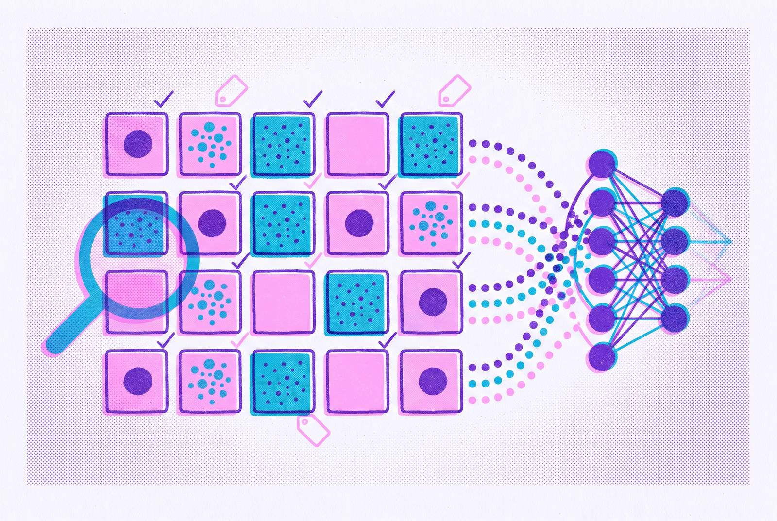
In supervised learning, a model learns pattern recognition from labeled examples. The images supply the features, while their labels supply the answers the model learns to predict. For reliable generalization, the model needs to see enough of those features, in enough different forms, during training. Dataset selection therefore makes clinical choices before a model ever assesses a patient. Which diseases appear, how they are photographed, and who names them all shape the resulting tool.
Architecture cannot fill gaps in what the training data show. A model trained mostly on dermoscopic images of neoplastic lesions in fair-skinned patients has not learned what it needs to assess smartphone photographs of inflammatory conditions in darker skin. The disease, image type, and patient population have all changed. This is why dataset composition is more than an engineering concern. It defines the clinical territory in which the platform has learned to operate.
The canonical public datasets that trained most of what is deployed today
HAM10000 contains 10,015 multi-source dermoscopic images covering seven pigmented lesion types, including melanoma, melanocytic nevus, basal cell carcinoma, actinic keratosis and intraepithelial carcinoma, benign keratosis, dermatofibroma, and vascular lesions. ISIC 2019 is a large curated image collection that, together with HAM10000, forms the backbone of competitive benchmarking in neoplastic skin AI. These collections gave researchers a shared basis for comparing lesion classifiers. Their prominence also helped make this particular clinical task the field’s standard test.
Other datasets reflect different choices about what needs documenting. Fitzpatrick17k contains tens of thousands of images with explicit skin tone annotations, built to introduce diversity lacking in ISIC-derived datasets. SCIN, developed by Google Health and Stanford Medicine in 2023, contains crowdsourced cases with one to three photographs, self-reported demographic data, and review by US board-certified dermatologists. It reflects the consumer-submitted photo context of telehealth. Xiangya Derm, from Xiangya Hospital, offers a large-scale clinical image collection spanning over 500 skin disease types.
These datasets do not measure the same capability. Their acquisition methods, diagnostic categories, and lesion labels differ, so results cannot be compared as though every model sat the same examination. A broad collection of clinical photographs asks something different from a focused dermoscopic benchmark. When a platform cites a familiar dataset name as evidence of clinical breadth, establishing its complete training history requires evidence beyond public benchmark use.
What "labeled" means in practice and why annotation methodology shapes what a model learns
A label defines what counts as the right answer. With one diagnosis per image, the model learns to classify the image into a named category. Rich text tied to clinical context teaches the model in a different way by linking what is visible with symptoms, history, and possible diagnoses. Two datasets of the same size can therefore lead models to behave differently in clinical use.
Derm1M makes that distinction concrete through an expert ontology. It covers 390 skin conditions with four hierarchical levels and 130 clinical concepts, rather than relying only on a flat list of disease names. Its contextual information includes skin tone, medical history, and symptoms. That structure supports learning relationships used in differential diagnosis. It gives a vision-language model more to work with than a single category, though richer annotation is not itself proof of sound clinical reasoning.
The source of the label also shapes its meaning. SCIN uses one to three dermatologists per case, allowing variation between readers to reflect some of the uncertainty present in clinical assessment. Single-expert labeling can look cleaner while encoding one clinician’s biases. Imaging modality adds another constraint because a label attached to a dermoscopic view does not make that view interchangeable with a clinical photograph. Expert annotation, sometimes requiring biopsy confirmation, is costly, which helps explain the repeated use of a small set of established datasets.
The structural bias baked into the dominant datasets and what it costs clinically
Lighter skin, dermoscopic acquisition, and neoplastic conditions receive much more attention than many other combinations of patient, image, and disease. HAM10000’s heavy skew toward fair-skinned individuals illustrates the consequences. In the comparison described in the research, melanoma-detection sensitivity falls from a functional level on HAM10000 to 11% on the Pipsqueak darker-skin dataset, indicating a failure to detect disease.
Adding skin tone labels does not automatically balance a collection. Fitzpatrick17k still has many times more samples of its most represented skin type than of its least represented type. SkinMap maps the aggregate public image corpus and finds that darker skin tones, Fitzpatrick V–VI, make up only a small fraction of images. Pediatric patients are similarly sparse. These gaps leave models with fewer examples to learn from, even when the total number of images in the field looks large.
Darker-skinned patients have lower melanoma rates but higher mortality from it, largely because of late diagnosis. Less diverse training data can contribute to delayed or incorrect diagnosis and widen health disparities. Patients with darker skin are also less willing to accept AI-assisted dermatology, which makes it harder to put these tools to use. Simply making a collection larger, including through synthetic data, would not by itself demonstrate that the missing clinical groups were adequately represented or that performance had improved for them.
Why distribution shift — not skin tone alone — is the deeper failure mode
Distribution shift changes what a benchmark result can tell the reader. Research decomposing performance gaps found that training on one clinical distribution and testing on another dominated across all evaluated models. A model may have learned a narrow combination of diseases, imaging methods, and populations that differs substantially from the cases it receives in practice. High accuracy within that combination does not establish accuracy outside it.
The benchmark-heavy core of the field emphasizes dermoscopic images of neoplastic conditions. Yet inflammatory, infectious, and everyday conditions such as acne, eczema, rosacea, psoriasis, and contact dermatitis account for most real-world dermatology volume. These conditions are sparsely represented in the benchmarks used to evaluate many models. SkinMap makes the mismatch visible through dense clusters of common neoplasms on fair skin and sparse coverage of underrepresented skin types and many rare diseases.
More neoplastic examples can improve neoplasm classification without building the condition recognition needed for telehealth triage. An ISIC benchmark score may therefore say little about assessing a smartphone photograph of a rash submitted through an asynchronous consultation. A high score is useful only when the evaluation also fits the care setting where the tool will be used.
How the field is responding with foundation models and million-scale vision-language datasets
PanDerm addresses the modality gap through broader pre-training. Published in Nature Medicine in June 2025, it was pre-trained through self-supervised learning on millions of real-world skin disease images from 11 clinical institutions across 4 imaging modalities. Its design moves beyond learning from a single benchmark collection. However, self-supervised pre-training is not the same as supervised training on millions of labeled patient cases.
Derm1M expands the image-text route. An ICCV 2025 Highlight publicly released in October 2025, it is 257 times larger than SkinCAP, the prior largest vision-language dermatology corpus. DermoGPT, a January 2026 preprint, is an open-weights and open-data model for morphology-grounded dermatological reasoning. It extends this approach toward interpretable clinical outputs.
These developments address different parts of the same constraint. Vision-language systems can generate differential diagnoses in clinical language, while self-supervised learning allows models to learn from unlabeled images before fine-tuning on labeled sets. Haggerty and Chandra show that targeted self-supervised pre-training on task-specific data can match large-scale self-supervised pre-training on ImageNet when labeled examples are scarce. SkinMap supplies a way to audit whether new collections expand clinical coverage or reinforce old gaps. None of these contributions alone establishes which model is best for every skin assessment task.
What human-AI collaboration looks like in practice and why clinician oversight is not optional
In PanDerm’s human-AI collaboration study, clinicians used model output to help assess cases. Clinicians first assessed cases independently and supplied their top-3 diagnoses. They then reviewed the model’s top three predicted diagnoses and could maintain or modify their original diagnosis and differential diagnoses. The clinician retained the decision-making role throughout the process. This workflow gives AI a defined purpose without treating its output as the final clinical answer.
Oversight addresses risks that broader training has not removed. SkinGPT-4 shows how a single model that covers many skin disease categories can be less precise with rare or underrepresented conditions. LLM-based systems without expert validation also risk hallucinated, overconfident, and clinically unsafe outputs. Those risks remain even when the clinical language reads well. Data on what patients prefer show that a strong majority trust dermatologist-guided AI, while almost no patients trust AI apps that work alone.
Triage makes the workflow especially consequential for access to care. In teledermatology, patients submit images and an algorithm assesses whether to escalate the case to a dermatologist, placing distribution mismatch at a decision point in care. AI Labs Group S.L., working with Puerta de Hierro University Hospital, studied a collaboration model prospectively in a clinical trial that has now ended. Its completion should not be confused with a reported positive result. The strongest case for AI-assisted triage remains a first step that extends specialist reach, with licensed clinician oversight integrated into the workflow.
What patients and clinicians should understand about reading a platform's capability claims
The practical lesson is to ask about clinical fit. Which conditions appear in the training data, and in what volume? A large collection concentrated on neoplasms offers limited evidence for assessing inflammatory or infectious conditions. Patients and clinicians should also check the imaging modality and whether the training photographs look like those the patient will submit. Evaluation on the intended kind of image is more informative than a strong score from a different setting.
What representation does the training set provide for Fitzpatrick V–VI skin tones, and how was performance evaluated for those patients? Missing evidence is not a basis for assuming equal reliability. Patients and clinicians can also ask who supplied the labels and what annotation protocol they followed. Expert-reviewed crowdsourcing, single-expert diagnosis labels, and rich clinical text each change what a model learns and what remains uncertain.
Finally, the assessment needs a clear place in care. Does a licensed clinician review the output before it reaches the patient, or does the tool present an AI answer directly with only a disclaimer? Those are structurally different services, even if both describe themselves as multimodal clinical AI trained on labeled dermatology cases. Used at the right step, AI-assisted assessment can help extend access to clinical expertise while leaving diagnosis and judgment where they belong.
Sources
- Frontiers
- A Global Atlas of Digital Dermatology to Map Innovation and Disparities
- Self-supervised learning for skin cancer diagnosis with limited training data
- A multimodal vision foundation model for clinical dermatology
- Derm1M: A Million-scale Vision-Language Dataset Aligned with Clinical Ontology Knowledge for Dermatology
- Exploring the Diagnostic Capability of Artificial Intelligence in Dermatology for Darker Skin Tones: A Narrative Review
- Benchmarking Off-the-Shelf Multimodal AI Models Against Dermatologists on Patient-Captured Skin Images