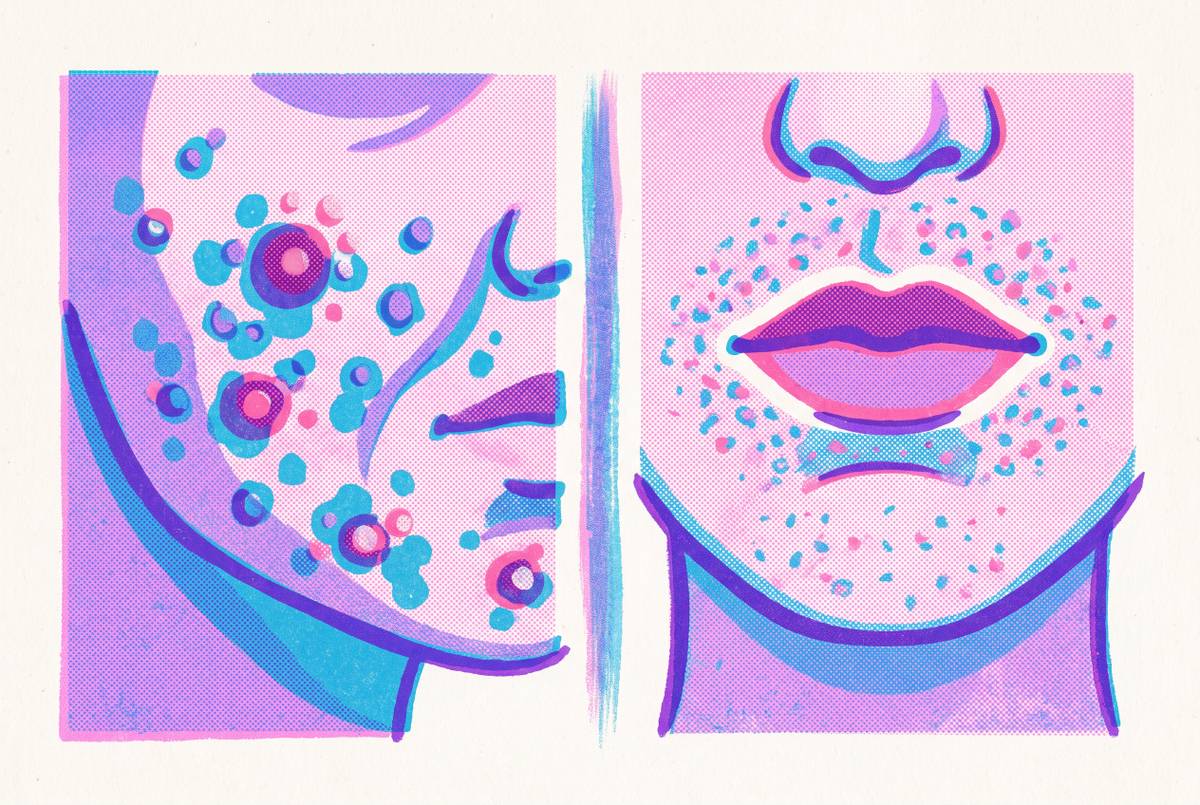
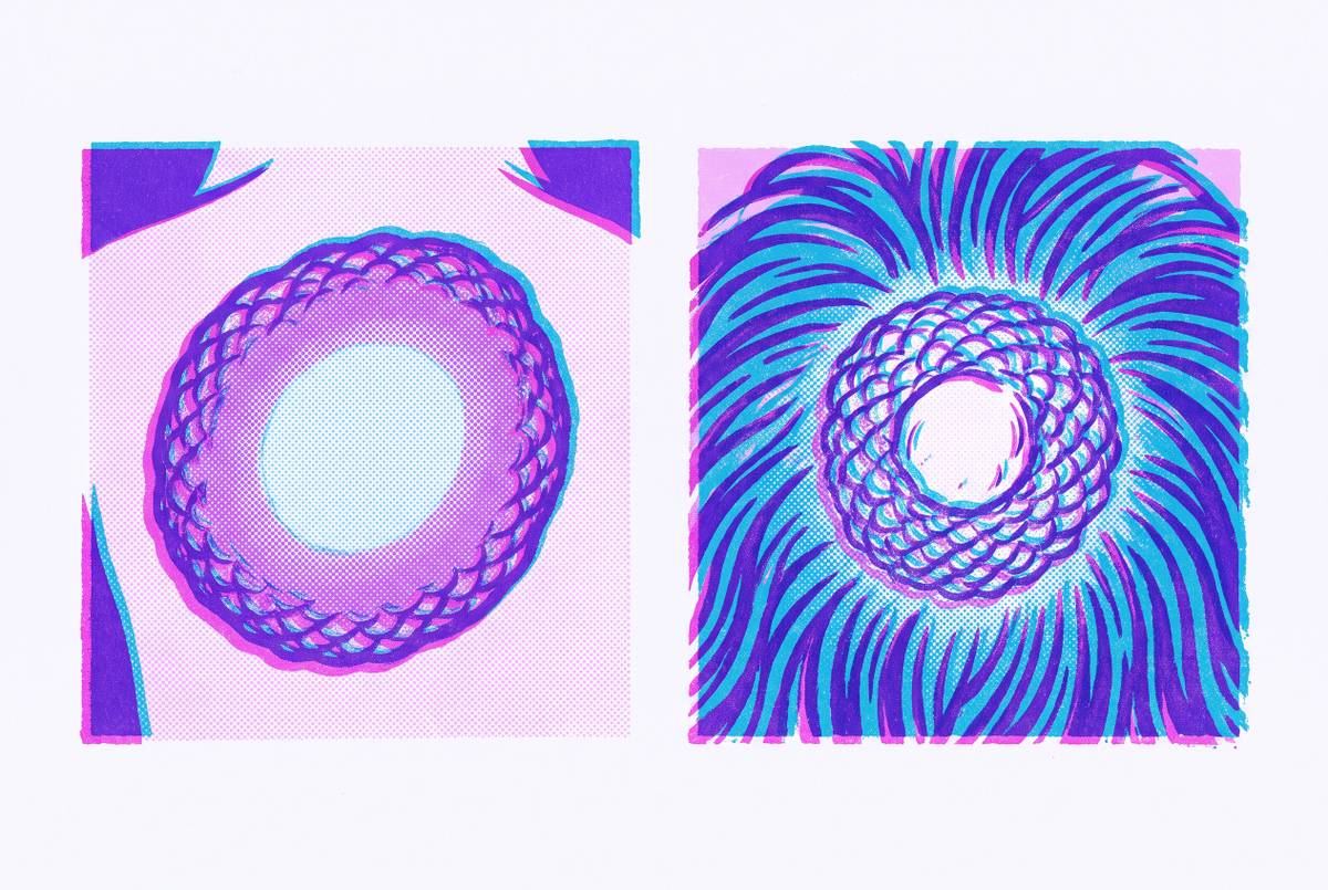
Milia Under the Eyelid Versus Other White Bumps
How to tell milia apart from four other white eyelid bumps.
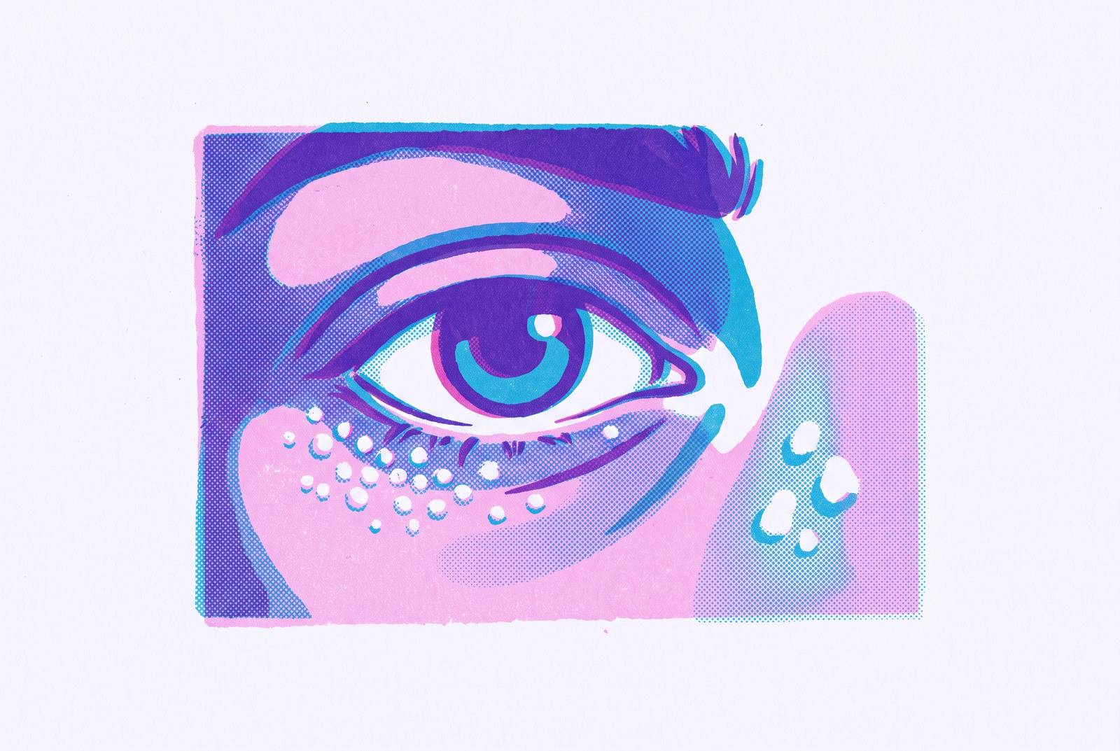
The skin on the eyelid is roughly 40% thinner than skin elsewhere on the face, and it carries far fewer sebaceous glands to keep it naturally exfoliated. That combination, thin tissue with almost no oil to smooth over rough patches, turns out to be exactly the wrong environment for keeping different biological processes separate. Keratin trapping, sweat duct overgrowth, and cholesterol deposition are three completely unrelated mechanisms, yet all three find a home in this same narrow strip of skin and all three produce small, pale bumps that look like cousins of each other. Milia, syringomas, xanthelasma, even the occasional flat wart, they can all sit within a few millimeters of each other and pass as the same thing. They are not, because the similarity in appearance masks distinct underlying conditions. Because the bumps cluster in the same locations and are similar in size (mostly 1–3 mm), visual inspection alone, without knowing what to look for, almost always produces a wrong guess.
What milia are: trapped keratin, not clogged pores
Start with the one everyone reaches for first, because milia is the default guess whenever a small white bump appears near the eye. Keratin is the structural protein that makes up skin, hair, and nails. When dead skin cells fail to shed the way they normally would, keratin builds up and gets sealed underneath the surface, forming a firm little dome with no visible opening.
That detail, no opening, means there is nothing to squeeze and no channel to drain, so trying anyway just tears skin that's already thin and risks scarring or infection. A whitehead has a pore you can work with. Milia doesn't. There's nothing to squeeze, no channel to drain. Trying anyway just tears skin that's already thin and risks scarring or infection, especially this close to the eye.
Milia break into two mechanistically distinct subtypes. Primary milia occur spontaneously, dead cells getting trapped at the base of a hair follicle or a sweat gland, and this is the version most commonly found on eyelids. Secondary milia are a response to something else entirely: trauma to the skin, burns, dermabrasion, long stretches of topical steroid use, or certain blistering skin conditions that leave keratin nowhere to go. UV damage plays a role too, thickening the outer skin layer just enough to slow normal shedding, and so do heavy, occlusive eye creams loaded with lanolin or mineral oil. Milia carry zero systemic implications, and that sets up a contrast that matters later. They're a nuisance of skin biology, not a signal about anything happening elsewhere in the body.
Visually, milia present as white to yellowish-white, firm to the touch, about 1 to 2 millimeters, with a smooth surface and no inflamed base. They show up scattered or in small clusters, and they don't seem to prefer any particular spot on the lid or under-eye area. Keep that image in mind. Everything else in this piece gets measured against it.
The three conditions most commonly mistaken for milia, and the one biological clue that separates each
Three other conditions get mistaken for milia often enough that it's worth walking through each one on its own terms (thin skin, minimal oil, and low friction create the right environment for keratin trapping, sweat duct overgrowth, and cholesterol deposition, three entirely separate biological mechanisms that all produce small pale bumps in the same small zone). Each has a distinct cause. Each leaves a different fingerprint.
Syringomas come from an overgrowth of cells inside the eccrine, or sweat, glands. That's a proliferative process, cells multiplying, distinct from the trapping process seen in milia. They show up more in women, particularly middle-aged women of Asian descent, tend to first appear during adolescence, and are also documented more frequently in patients with Down syndrome. Purely a cosmetic concern, nothing more.
Xanthelasma changes the conversation. These are soft, flat, yellowish-orange plaques built from cholesterol-laden immune cells that gather beneath the eyelid's thin skin, usually near the inner corners. The color alone should be the first flag: yellow-orange, not the white-pearl of milia.
But the real reason xanthelasma deserves more attention than a bump this size would normally get: roughly half the patients who have it also show elevated cholesterol, triglycerides, or other lipid abnormalities, and xanthelasma itself shows up in an estimated 0.3 to 1.1 percent of the general population. That's not a number to write off, even for someone who feels perfectly healthy. Researchers at Massachusetts Eye and Ear presented data at the 2025 annual meeting of the American Academy of Ophthalmology showing a measurable link between xanthelasma and major cardiovascular events like heart attack and stroke. So a bump that looks purely cosmetic can, in effect, be a signal flare for something happening in the cardiovascular system. A yellow patch near the eye might be worth a phone call to a primary care doctor, not just a dermatologist.
Then there's the odd one out. Flat warts, or verrucae planae, come from human papillomavirus, specifically HPV types 3, 10, and 28. Every other condition in this piece traces back to trapped protein or overgrown cells. This one is viral, a completely different biological story. Flat warts can look flesh-colored or slightly yellow, flat, smooth, easy to mistake for milia at a glance. But because the cause is viral, treating it like milia (retinoids, extraction) won't do anything. The mechanism doesn't match the tool. The distinguishing features are flesh-toned to slightly yellowish, firm, 1–3 mm bumps appearing predominantly on the lower eyelids, often bilaterally and in groups. The key visual clues are the yellow-orange color (distinct from the white-pearl of milia), softness rather than firmness, flat rather than dome-shaped, and a notably larger size (ranging up to 30 mm) versus milia's 1–2 mm.
Four descriptors that let a patient distinguish the most likely culprit before seeing a clinician
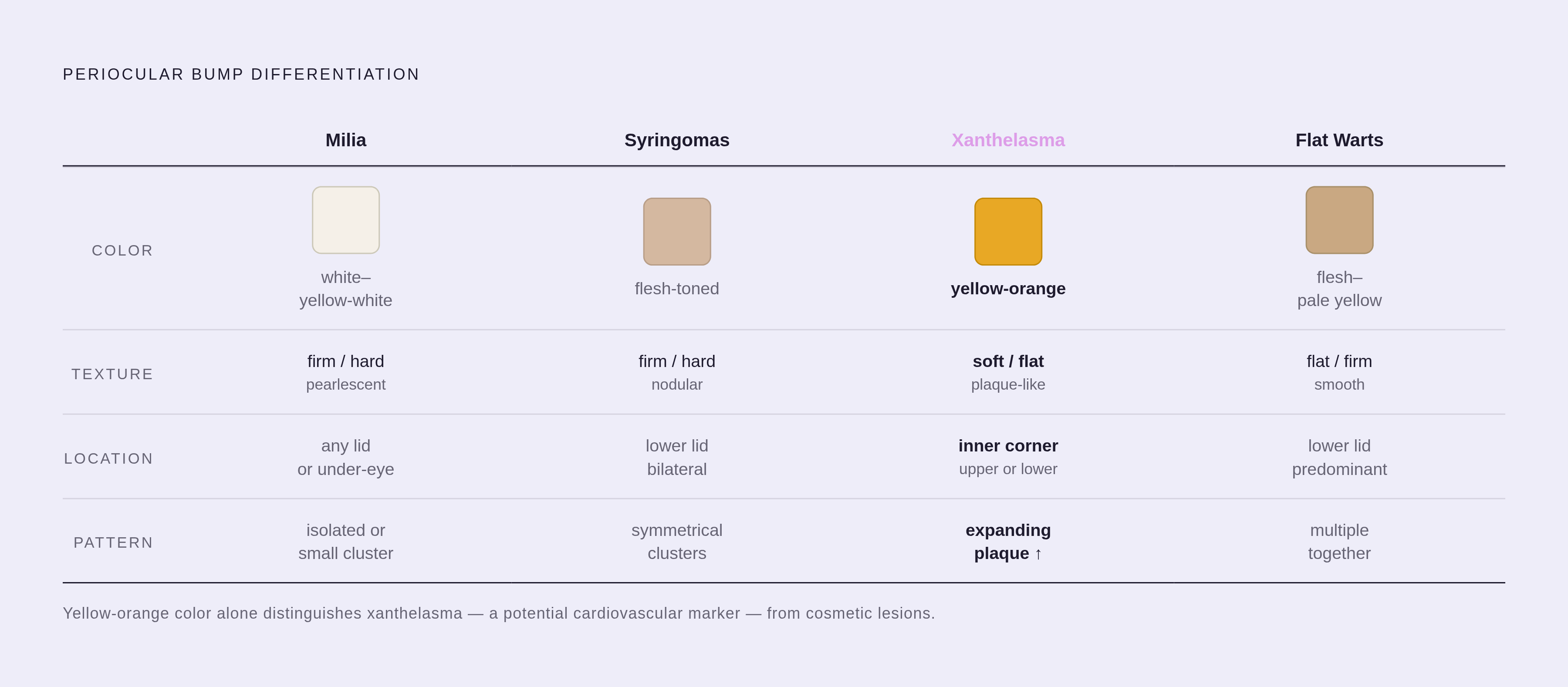
So how does someone actually use all this before booking an appointment? Four things to check: color, texture, location, and pattern. These are the same four things a clinician is going to ask about anyway, so gathering them ahead of time isn't just useful, it's the exact information a remote visit or an in-person exam will need first.
On color: white to yellowish-white and pearl-like points toward milia. Flesh-toned blending into the surrounding skin suggests syringoma. Yellow-orange, distinctly different from the skin around it, suggests xanthelasma. Flat and skin-colored leans toward a wart.
On texture, hard and firm covers both milia and syringoma, so texture alone won't separate those two. Soft and flat is xanthelasma's territory.
On location, milia can show up anywhere on the lid or under the eye, scattered without much pattern. Syringomas cluster on the lower eyelid, usually on both sides. Xanthelasma tends to sit near the inner corner of the upper or lower lid.
On pattern and onset, something that grew in slowly, sits isolated or in a small cluster, and doesn't hurt points to milia or syringoma. A flat plaque that's been slowly expanding, painless, suggests xanthelasma. Multiple flat bumps appearing together in a group should put flat warts on the list.
None of this replaces an exam. But a few danger signals override the whole framework and call for prompt in-person evaluation rather than a wait-and-see approach: a bump that bleeds repeatedly or ulcerates, a pearly or waxy border that looks rolled at the edges, lost eyelashes near the bump, any distortion of the lid margin, a translucent or bluish cyst, or a firm non-tender nodule sitting deeper in the lid tissue away from the lash line. A tender red bump right at the lash line with a visible pustule is its own thing too, likely a stye, and belongs to a separate differential. The full list of what a periocular bump could turn out to be also includes trichoepitheliomas, papular sarcoidosis, and basal cell carcinoma, none of which anyone should try to sort out solo.
Whether a photo-based telehealth visit can reliably assess a white eyelid bump
Once someone has gathered those four descriptors, the next question is practical: can this actually get sorted out over telehealth, or does it need an in-person look? The honest answer sits in the middle. A systematic review found teledermatology reaches meaningful, though imperfect, agreement with face-to-face evaluation overall, and that agreement climbs noticeably when patients get real guidance on how to take the photo, lighting, angle, how close to get. A systematic review and meta-analysis out of Semmelweis University, published in Frontiers in Medicine, went further, concluding that teledermatology shows high diagnostic accuracy and holds up as a reliable alternative to in-person care for general skin conditions.
That's an encouraging baseline. But eyelid bumps sit in an odd spot within that data, because the conditions that matter most here are also the hardest to photograph well.
Classic milia photograph fine. The white-pearl color and the firm, domed shape tend to come through clearly in a decent close-up under good light. Syringomas usually photograph well too, since their location on the lower lid and their symmetrical pattern are visible traits that don't depend much on subtle color rendering.
Xanthelasma is the harder case. Its yellow-orange hue is subtle, and phone cameras under typical indoor lighting can wash that color out or shift it just enough that a reviewing clinician misses the visual cue that would normally prompt a lipid panel referral. And any lesion with a pearly, waxy look, or one that bleeds on and off, needs an in-person look, full stop. Basal cell carcinoma sits in the same differential as everything else discussed here, and no photo review should be the final word when that's on the table.
Some telehealth platforms now run AI-assisted triage ahead of clinician review. A 2024 proof-of-concept study in the European Journal of Dermatology tested an AI-assisted tele-expertise platform and found its top-3 algorithm hit high sensitivity and specificity for common dermatoses, useful for flagging patterns and organizing lesion characteristics before a person looks at the case. Most current AI classifiers in dermatology are built around morphology-based neoplasm detection, which leaves a lot of inflammatory and history-dependent presentations, periocular conditions very much included, underserved by the algorithm. Human oversight isn't optional here. The eyelid's differential runs from a totally benign keratin cyst all the way to carcinoma, and that range is too wide for pattern-matching software to close alone.
Practically, that means asynchronous telehealth (sending photos and a description for a clinician to review) is a reasonable starting point for a bump that fits the milia or syringoma profile and shows none of the danger signals covered earlier. It is not the right starting point for anything showing those signals, or for a bump the patient genuinely can't characterize with any confidence.
Treatment approaches matched to the condition
Everything above sets up the actual stakes: get the identification wrong, and the treatment goes wrong with it.
For milia, patience is often the first move. Primary milia frequently clear on their own over weeks to months, and in infants, they often resolve within a few weeks without intervention. For milia that stick around, topical retinoids like tretinoin, applied nightly, speed up cell turnover and can gradually clear them out. A comprehensive review covering 23 randomized controlled trials found solid evidence behind isotretinoin, retinol, and tretinoin for improving skin texture generally. Professional extraction is the fastest fix: a clinician uses a sterile needle or blade to create a tiny opening and expresses the trapped keratin out. For milia that persist or spread, dermabrasion, chemical peels, microdermabrasion, or erbium:YAG laser ablation are all options, though each carries more risk this close to the orbital rim and belongs in a professional's hands, not a bathroom mirror. Squeezing or lancing milia at home near the eyelid is a bad idea regardless of how tempting it looks in the mirror. It's thin skin over a delicate structure, and infection or scarring is a real possibility.
One caution on retinoids specifically: they increase photosensitivity, they're not considered safe during pregnancy, and using them around the eyelid deserves extra care given how thin that skin already is. Some evidence suggests retinoid use near the eyes may contribute to dry eye disease, so getting professional guidance before starting a periocular retinoid routine is a reasonable step, not an overcautious one.
Now flip the scenario. Someone with xanthelasma who assumes it's milia and starts applying retinoid cream nightly wastes product while delaying the conversation that actually matters: a fasting lipid panel and a talk with a primary care doctor about cardiovascular risk. That's the real cost of misidentification here. It's a missed cosmetic-level judgment that carries real medical stakes. It's a missed signal.
Syringomas don't respond to milia treatments at all, since the mechanism is entirely different, an overgrowth of gland cells rather than trapped keratin, so retinoids and needle extraction aren't built for that job. And flat warts, being viral, need an approach aimed at the virus itself, not cell turnover or manual extraction.
The throughline across all of it: four bumps, four different causes, and only one of them, xanthelasma, comes with a health question attached that reaches well beyond the eyelid. Knowing which one is looking back in the mirror is about picking the right cream and about knowing when a bump is asking for more than a cream. It's about knowing when a bump is asking for more than a cream.
Sources
- Milia Under Eye: Complete Treatment & Prevention Guide 2025
- Milia (Milk Spots): Causes & Treatment
- Flat Warts and Syringomas | Contemporary Pediatrics
- A Case Report of Xanthelasma and the Associated Differential Diagnosis
- Syringoma: What it is, vs Milia, Causes & Treatment
- Benign Eyelid Lesions - StatPearls - NCBI Bookshelf - NIH
- Image-based Telehealth Effective for Benign Eyelid Lesion Diagnosis