Skin Conditions That Signal Systemic Disease

The skin is the only internal organ you can see without a single instrument. That fact alone makes it one of the best early warning systems the body has, and after years of watching patients and clinicians catch (or miss) that signal, I've come to think we undervalue it. Six organ systems in particular, the hepatic, renal, gastrointestinal, hematological, cardiopulmonary, and neurological, tend to write themselves onto the skin with real consistency. This piece is about learning to read that writing before it turns into something louder.
Here's the mechanism, stated plainly: the skin externalizes what's happening inside. Immune activity, vascular changes, metabolic shifts, inflammatory cascades, they all leave marks at the surface, sometimes months or years before a person feels sick anywhere else. The gut-skin axis is a good example of this in action: the gut and skin talk to each other constantly, and a shift in gut microbiome composition can alter systemic immune responses in ways that show up as a rash or texture change on the arm or face. A newer field called dermatogenomics, which pairs visible skin phenotypes with the genetic factors behind them, is pushing this even further. A January 2026 paper in the MDPI journal DNA, out of the University of British Columbia, makes the case that combining what we see on skin with what we know about polygenic risk is opening the door to earlier disease recognition than either approach manages alone.
So here's the clinical implication, stated without hedging: a pattern on the skin is sometimes the first legible signal of a disease that hasn't produced a single other symptom yet, though not always. But often enough that learning the patterns matters.
How autoimmune disease announces itself on the skin before it damages organs
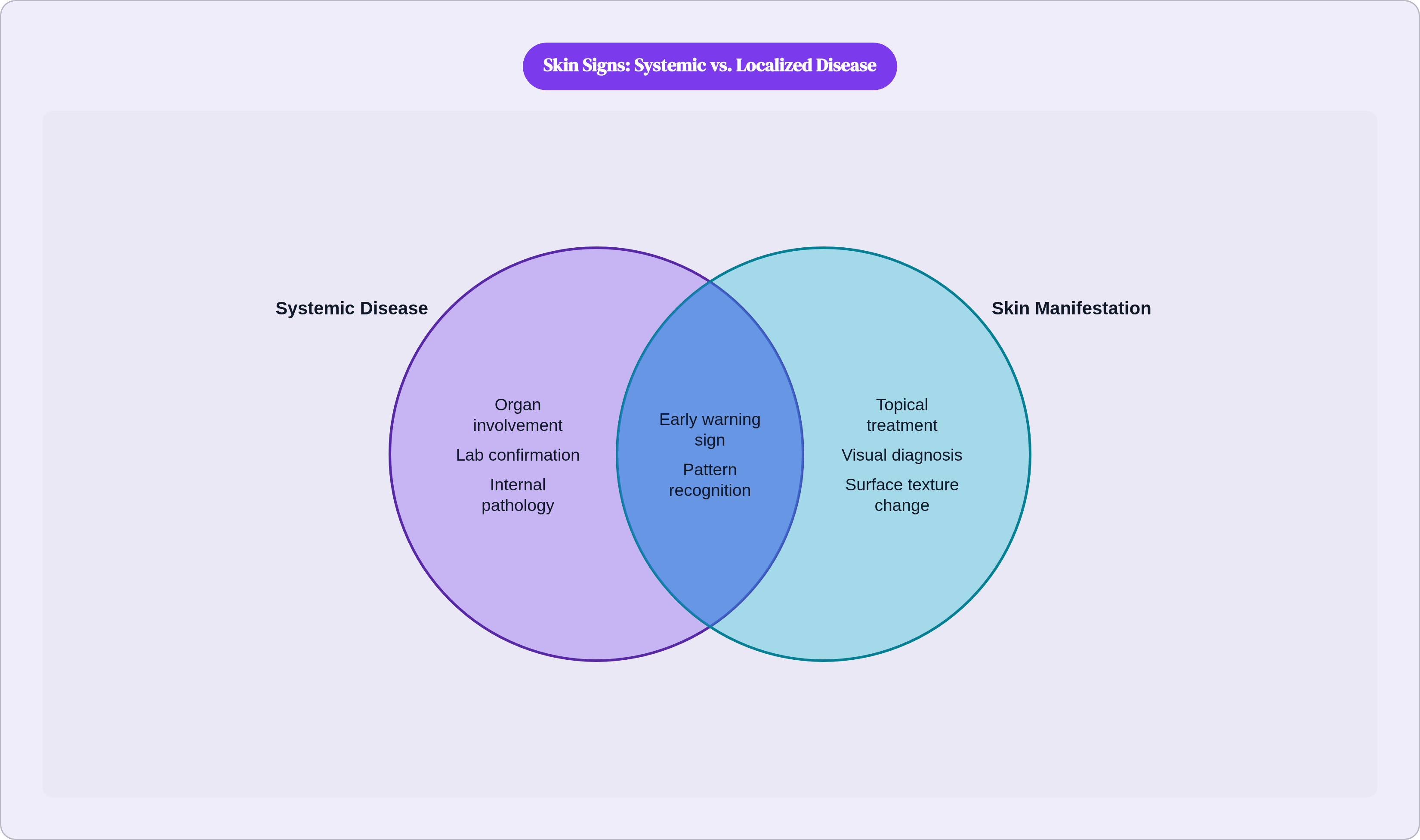
Take lupus. Up to 85% of people with systemic lupus erythematosus develop skin involvement at some point, and the malar rash, that reddish, slightly scaly patch across the cheeks and nose bridge that people call the "butterfly rash," is frequently the very first sign anyone notices. Lupus hits women roughly nine times more often than men, and catching that rash early carries real weight, because the disease doesn't stop at skin, going after kidneys, heart, and brain instead. Worth knowing too: UV exposure, infections, and stress can trigger flares in people who are genetically predisposed, which means a patient who notices their rash gets worse after a weekend at the beach is handing their doctor a genuinely useful clue.
Dermatomyositis works on a similar principle but with its own signature. A heliotrope rash (a violet discoloration across the upper eyelids, sometimes puffy) paired with Gottron's papules over the knuckles is close to diagnostic on its own. Add the shawl sign across the neck and upper back, the V-sign across the chest, and abnormalities at the nail fold, and you've got a pattern that's hard to mistake for anything else. What's striking is the sequence: these skin findings often show up before the muscle weakness does. A 2026 review in the MDPI Journal of Clinical Medicine notes they can be sufficient for diagnosis even in cases where muscle involvement never fully develops, the so-called amyopathic form. And there's a cardiac angle here that doesn't get enough attention: dermatomyositis can trigger myocarditis, and it shows up alongside lupus, rheumatoid arthritis, scleroderma, and Sjögren syndrome often enough that clinicians watch for overlap.
Then there's scleroderma, where the body overproduces collagen and the skin tightens and hardens as a direct, visible result. The same fibrotic process doing that to the skin is doing it internally, scarring the gastrointestinal tract, the lungs, the cardiovascular system. Flags to watch for: skin thickening, puffy or swollen fingers, stiffness in the hands, painful ulcers at the fingertips, with Raynaud's phenomenon and GERD tending to travel alongside them. And dermatitis herpetiformis, an intensely itchy, blistering rash linked to celiac disease, is one more case of a gut disorder writing its symptoms onto skin instead of staying quietly internal.
Notice the thread running through all three of these. The skin sign shows up early, you can see it without ordering a single test, and it points toward a specific organ system. That's not incidental; that's pattern recognition doing real clinical work.
What psoriasis and acanthosis nigricans reveal about cardiovascular and metabolic risk
Psoriasis affects somewhere around 2% to 3% of the population, and for a long time it got filed under "skin problem" and left there. That framing is outdated. Psoriasis is now understood as a systemic inflammatory disease, and people who have it are up to 50% more likely to develop cardiovascular disease than the general population, with the risk climbing as the skin involvement gets more severe.
There's a model for this called the "psoriasis march": systemic inflammation combines with obesity, which drives insulin resistance, which damages the blood vessel lining, which paves the way for atherosclerosis. It's a chain, not a coincidence. A 2025 study in JAMA Dermatology (161(1):81-86) used mediation analysis, a statistical method for testing whether one factor actually explains the path between two others, and found that systemic inflammation, measured through a marker called GlycA, partially explains the link between how severe someone's psoriasis is and their cardiovascular outcomes. That's not a loose association anymore; that's a mechanism with numbers behind it.
It goes deeper than inflammation, too. A May 2025 paper in Nature Reviews Cardiology found that psoriasis and atherosclerotic cardiovascular disease share genetic loci, including PCSK9 and APOE, genes with well-established roles in cholesterol metabolism and cardiovascular risk. The connection is partly baked in at the genomic level, before inflammation even enters the picture. Which raises a fair question: if the risk is partly genetic and partly inflammatory, what should a clinician actually do differently at diagnosis? The 2025 PPACMAN consensus has an answer. It recommends baseline cardiovascular risk assessment the moment someone is diagnosed with psoriasis, especially for moderate-to-severe cases or anyone who'll need biologic therapy, along with routine screening for hypertension, diabetes, dyslipidemia, obesity, and metabolic syndrome.
Acanthosis nigricans tells a related but distinct story: insulin resistance made visible. The appearance is unmistakable once you've seen it: velvety, darkened, thickened skin in the folds, armpits, groin, back of the neck. Per DermNet NZ, reviewed in 2025, it's almost always a sign of an underlying metabolic or hormonal issue, most commonly type 2 diabetes or insulin resistance. And the prevalence numbers say a lot about who's carrying the heaviest metabolic burden: under 1% in Caucasian populations, 5.5% among Latino populations, 13.3% among African Americans, and 34.2% among Native Americans in the United States. The mark itself isn't the disease. It's a pointer, and a genuinely useful one, because it can flag a metabolic process worth addressing before it becomes frank diabetes.
Other metabolic-skin links deserve a mention here too. Acne has been tied to insulin resistance and a cellular growth pathway called mTORC1 activation, while hidradenitis suppurativa travels with obesity and metabolic syndrome. And androgenetic alopecia, ordinary pattern hair loss, has been connected to insulin resistance as well, according to a June 2025 review in Forum Dermatologicum. None of these are standalone cosmetic issues once you look at what tends to sit underneath them.
Paraneoplastic skin findings and how cancer signals itself before a diagnosis is made
Paraneoplastic dermatoses are skin disorders that show up alongside an internal cancer, not because tumor cells have invaded the skin, but because the tumor is sending out hormonal or immune signals that land somewhere else in the body. The clinical significance is real: per UpToDate, when one of these conditions appears before the cancer itself has been found, recognizing it can be what prompts the workup that finds the tumor, or flags a recurrence early after treatment.
Paraneoplastic pemphigus is the most severe example. It's uncommon, but it's a life-threatening mucocutaneous syndrome, and its defining feature is severe stomatitis, inflammation and sores in the mouth, that doesn't respond to standard treatment. It shows up frequently alongside non-Hodgkin lymphoma and chronic lymphocytic leukemia. The most dangerous complication, per a 2023 paper in Frontiers in Medicine, is bronchiolitis obliterans, a scarring of the small airways that can be fatal.
Glucagonoma syndrome and carcinoid syndrome work differently: these are tumors that secrete hormones, and those hormones produce their own characteristic skin changes as a downstream effect. Sweet's syndrome and pyoderma gangrenosum, both inflammatory skin conditions, can also point toward an underlying blood cancer.
It's worth drawing a clear line here between two very different pathways. One is direct metastasis, cancer cells actually infiltrating the skin, and the other is the paraneoplastic effect, where the tumor is somewhere else entirely and it's hormonal or immune signaling doing the damage at the surface. These carry different clinical urgency, and conflating them does readers a disservice. None of this means a given rash confirms cancer is present. These are signals that warrant evaluation, not diagnoses on their own, and that distinction matters more than almost anything else in this section.
Hepatic and gastrointestinal disease and the skin changes that accompany them
Generalized itching with no rash to explain it, what clinicians call pruritus, is one of the most well-documented, non-specific skin signals of liver trouble, including conditions like primary biliary cirrhosis. The mechanism involves bile salts building up in the bloodstream and interference with the body's own opioid signaling. It's an odd one because there's often nothing to see, just the itch itself, which is exactly why it gets missed or dismissed.
Other hepatic signs are more visible: jaundice, the yellowing of skin and eyes; spider angiomata, small clusters of blood vessels that radiate out from a central point like a spider's legs; palmar erythema, a reddening of the palms; and xanthomas or xanthelasmas, fatty deposits that show up with cholestatic liver disease and high cholesterol.
On the GI side, dermatitis herpetiformis comes back into the picture here as much a gut signal as an autoimmune one, since it's directly driven by gluten sensitivity and tied to celiac disease. Inflammatory bowel disease has its own skin companions too: pyoderma gangrenosum and erythema nodosum show up in a meaningful share of IBD patients, and they can appear before bowel symptoms flare or run in parallel with them.
Rheumatoid arthritis is worth bringing in here as well, even though it's usually filed under joint disease. It produces subcutaneous nodules, cutaneous vasculitis, and panniculitis, inflammation of the fat layer under the skin, which is a good reminder that these organ-system categories are porous, since disease doesn't respect the chart headings we use to organize it.
All of which loops back to that gut-skin axis mentioned earlier. The signaling between gut and skin microbiota runs both directions, and a shift in gut microbiome composition can alter systemic immune responses in ways that surface, quite literally, on the skin. It's the same mechanistic thread from the opening, just showing up again in a different organ system.
What distinguishes a skin change worth evaluating from normal variation
Not every rash means something is systemically wrong. I want to say that plainly, because the goal here isn't to have every reader convinced their dry patch is lupus. So what actually separates a skin change worth getting looked at from something that's just, well, skin being skin?
A few features tend to raise the stakes:
- Persistence. A change that doesn't resolve in a reasonable window, without an obvious cause like a new detergent or a sunburn, deserves a second look.
- Distribution pattern. Specific locations matter more than general ones. A butterfly pattern across the cheeks, flexural involvement in the armpits or groin, eyelid or knuckle changes, these carry more diagnostic weight than a generalized rash spread evenly across the body.
- Associated symptoms. Fatigue, joint pain, muscle weakness, unexplained weight change, or GI symptoms showing up alongside a skin change make the case for systemic investigation considerably stronger.
- Resistance to topical treatment. If a skin issue isn't responding the way it should to standard over-the-counter or prescribed topical care, that's a clue something underneath might be driving it.
Rapid change is its own category worth naming separately: skin that's spreading or evolving quickly, particularly in older adults, warrants prompt evaluation rather than a wait-and-see approach.
Here's something that doesn't get said enough: how well a patient describes what they're seeing directly affects how fast a clinician can connect the dots. Onset, location, color, texture, what makes it better or worse, that description is clinical data in its own right, not just a preamble before the real exam starts. At the SDPA Fall 2025 conference, clinicians were specifically urged to sharpen their recognition of rashes that might signal systemic autoimmune disease, and to get clearer on when to refer to rheumatology versus starting treatment themselves. Even trained dermatologists are working from pattern recognition here, not certainty, and that should tell you something about how much room there is for careful observation to matter.
How clinical evaluation of skin-systemic connections actually works today
In practice, the workflow usually starts with a visual assessment. A clinician looks at the skin, takes a history, and that combination steers what happens next, autoantibody panels, metabolic panels, imaging, or a referral. Dermatology increasingly sits at a boundary with internal medicine rather than inside a walled-off specialty, which is exactly the point being made at SDPA Fall 2025: recognizing when a skin finding needs rheumatology, oncology, gastroenterology, or cardiology involvement is now part of the job description, not an occasional exception.
But access is where this whole system tends to break down. Dermatology appointments can take weeks or months to land, and in that gap, a patient can sit with an unexamined skin change far longer than is comfortable, or safe. Early, low-barrier evaluation, someone reviewing a description or a photo of a skin finding, can do meaningful triage work: sorting which cases genuinely need urgent specialist attention from which ones just need monitoring.
This is where AI-assisted visual assessment earns its place, cautiously. Large multimodal models trained on extensive libraries of labeled clinical images can spot patterns in skin photos worth flagging for a clinician's review. The value there is speed and access, getting more eyes on more cases faster, not replacing the judgment a trained clinician brings to the table. Human oversight isn't optional in this loop; it's the whole point, because an AI flag means nothing until a licensed clinician looks at it, weighs the context, and decides what it actually means for that patient.
This is roughly the model Nolla is built around: AI-assisted image review paired with asynchronous clinician evaluation, giving patients a credible first look and a clear next step for the skin concerns that make up the vast majority of what people actually deal with, the ones that aren't emergencies but still deserve a real answer. It's designed around where the access gaps genuinely sit.
But I want to be honest about the ceiling here, because overselling this would undercut everything I just said about taking skin signals seriously. Telehealth and AI-assisted evaluation are a strong first step for pattern recognition and triage, though they are not a stand-in for an in-person workup once systemic disease is suspected and you need lab work or imaging to confirm it. Knowing the difference between those two moments, when a photo and a clinician's read are enough, and when you need to be in an exam room getting blood drawn, is arguably the most important skill this entire piece is trying to build.