Cellulitis Warning Signs That Need Prompt Medical Attention

Not every cellulitis case starts from the same baseline, and knowing where you stand changes how aggressively you should respond to early symptoms.
Conditions that impair immune function sit at the top of the list: diabetes, kidney disease, immunosuppression from medications or underlying illness. These aren't just comorbidities you mention once at intake. They are active variables in how fast an infection moves and how well your body can slow it down before intervention catches up.
Structural factors compound the picture. Lymphedema and chronic venous insufficiency create environments where fluid pools, circulation slows, and bacterial clearance stalls. Obesity belongs here too. A large-scale 2026 retrospective study identified leg ulcers and lymphedema specifically as key drivers of cellulitis incidence and complication risk, which tracks with what clinicians see repeatedly: the anatomical setup precedes the infection.
Then there are the skin conditions that keep creating entry points: tinea pedis, eczema, shingles. Each produces microbreaks in the cutaneous barrier that bacteria exploit, sometimes through skin that looks completely intact. No visible wound doesn't mean no entry point, and that distinction trips people up more than almost anything else in this condition.
Recurrence is its own concern. Annual recurrence rates for leg cellulitis run roughly 8% to 20%, typically in the same anatomical location. If you've had it before, your threshold for seeking care on a new episode should be lower, not higher. A presentation that looks mild in someone with lymphedema or diabetes is a moderate-risk presentation in clinical terms, regardless of how it appears on the surface.
One more thing before moving on: cellulitis of the face, genitalia, or the area around the eyes is high-risk even when symptoms appear superficially minor. Location does more work than visual severity in those regions, and proximity to critical structures is the variable that actually matters.
The Warning Signs That Mean the Infection Is Escalating
Redness that expands noticeably over hours is not something to monitor from your couch. Act on it immediately.
Red streaks extending outward from the infected area are a specific signal: lymphangitis, meaning the infection has begun tracking along the lymphatic channels. That is a qualitatively different situation than localized inflammation. The lymphatics are not supposed to be involved, and when they are, the trajectory changes.
Fever at or above 100.4°F alongside worsening local pain and swelling indicates a systemic response is underway. So do changes in skin texture beyond redness. Pitting or dimpling resembling an orange peel, blistering on the affected area: both are CDC-listed warning signs of escalating cellulitis. Skin turning purple or black means tissue is compromising, and that warrants emergency evaluation without delay.
Systemic signals (confusion, tachycardia, rapid breathing) shift the picture significantly. When these accompany local infection signs, you are approaching sepsis territory. The Sepsis-3 framework replaced the older SIRS-based definition after 2016, but the underlying logic holds: multiple systemic signals together represent a different emergency than a single elevated temperature. The older framework is gone; the clinical reality it was trying to capture is not.
Numbness in or around the infected area is easy to overlook and critically important. Loss of sensation suggests deeper tissue involvement. It gets misread as the infection quieting down when it is actually progressing.
The 48 to 72 hour checkpoint deserves particular attention. On oral antibiotics, cellulitis should begin showing measurable improvement within that window. If it hasn't, that absence of response is itself a warning sign, not a reason to extend the wait.
When the Location of the Infection Changes Everything
Periorbital cellulitis (infection in the tissue around the eye socket) requires immediate attention because of proximity to orbital structures and documented risk of vision complications if the infection advances. Orbital cellulitis, where the infection has moved behind the eye, is a frank emergency. Vision loss is a real outcome of delayed treatment here, not a theoretical one clinicians invoke to get compliance.
Facial cellulitis carries disproportionate risk relative to its appearance because venous drainage patterns in the face create pathways that infections elsewhere in the body simply don't have access to. What presents as moderate facial redness with some swelling is more immediately dangerous than a visibly severe presentation on your leg. The anatomy is doing something the appearance can't tell you.
The practical rule: facial swelling, swelling around the eye, or any visual disturbance alongside redness in those areas means an emergency department, not urgent care. "It doesn't look that bad" carries essentially no useful information when the face or orbital region is involved, because the risk there is driven by anatomy, not appearance. A telehealth session is also insufficient in those situations, for the same reason.
How to Tell Cellulitis from the Conditions That Look Like It
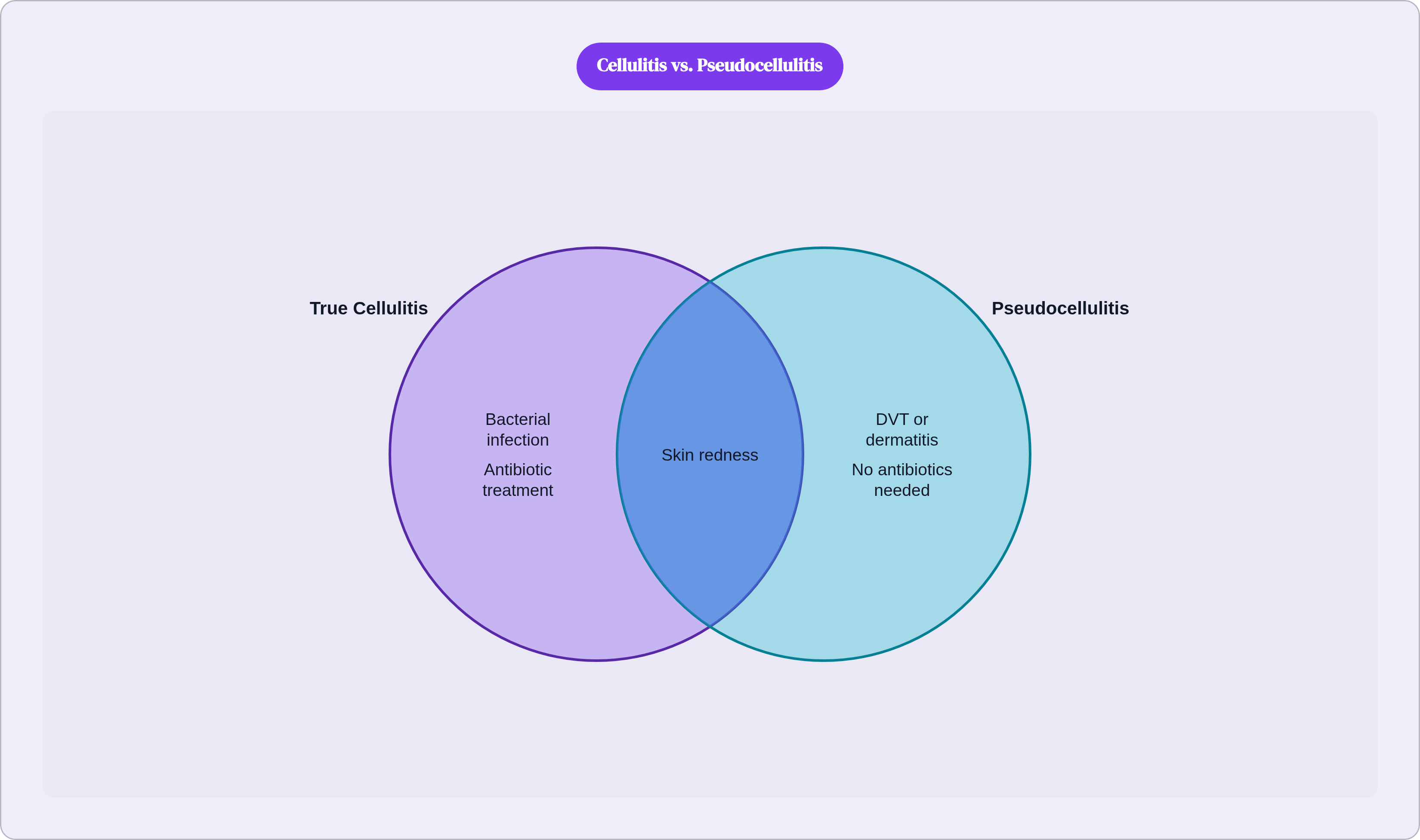
The misdiagnosis problem is substantial enough that it belongs in any serious conversation about this condition. Published studies report that between 28% and 33% of cases diagnosed as cellulitis are actually other conditions, a category clinicians call pseudocellulitis.
Common mimics include deep vein thrombosis, contact dermatitis, and shingles. Each requires a fundamentally different treatment approach, and the consequences of getting this wrong run in both directions. Patients with pseudocellulitis receive antibiotic courses they don't need: an estimated 44,000 such patients annually. The misdiagnosis also drives an estimated 50,000 to 130,000 excess hospitalizations per year and up to $515 million in avoidable healthcare expenditure. That's not a rounding error. That's a systemic diagnostic problem.
Dermatology consultation substantially reduces error rates. In one study, a dermatologist recommended discontinuing antibiotics in 82% of patients already prescribed them for presumed cellulitis, identified pseudocellulitis in 51% of admissions, and generated an estimated $200 million in annual savings attributable to specialist review. The numbers are striking precisely because the initial diagnoses were made by competent clinicians working from limited information.
What this means practically: a diagnosis that doesn't track with how your symptoms are evolving, or antibiotics that produce no meaningful improvement within 48 to 72 hours, are legitimate clinical reasons to request specialist review. It's a well-documented diagnostic problem, and the burden of flagging it often falls on the patient.
What Happens When Escalating Cellulitis Goes Unaddressed
The timeline compresses faster than most people expect.
Infection that isn't arrested can spread from soft tissue into the bloodstream, lymph nodes, bones, and cardiac tissue. Bone involvement produces osteomyelitis; bloodstream dissemination produces sepsis. Hopkins Medicine explicitly lists gangrene, amputation, and shock among the outcomes of untreated severe cellulitis. These are documented progressions, not cautionary embellishments.
Necrotizing fasciitis represents the most feared escalation. It is distinct from cellulitis, but it can be difficult to differentiate in early stages, and that ambiguity carries lethal stakes. The distinguishing features: rapidly advancing, non-blanching erythema; pain that is severe and disproportionate to what the skin looks like, followed paradoxically by reduced sensation as tissue necrosis progresses; hemorrhagic blistering; crepitus under the skin from gas-producing bacteria; early signs of septic shock or multiorgan dysfunction.
That sequence (severe pain followed by numbness) is the one to remember. The reduction in pain can be misread as improvement. It is not. By the time sensation diminishes, the process has already advanced significantly.
The window for acting on warning signs is measured in hours, not days.
Matching the Level of Care to the Level of Urgency
Emergency services or an ER immediately: facial or orbital involvement; skin turning purple, black, or blistering significantly; high fever combined with confusion, rapid heartbeat, or rapid breathing; red streaks spreading outward from the wound; any sign of septic shock. These are not presentations to observe overnight.
Same-day in-person urgent care: fever at or above 101°F; rapidly spreading redness or severe pain; pus, orange-peel skin texture, or early blistering; numbness in the affected area; high-risk patients with diabetes, immunosuppression, or lymphedema presenting with even apparently mild symptoms. The risk stratification for that last group starts higher than it does for someone without those factors, and that baseline matters more than the severity of what you can see.
Telehealth is appropriate for uncomplicated presentations when red-flag signs are absent. A licensed clinician can assess, prescribe, and establish a monitoring plan remotely when the clinical picture supports it. Nolla provides licensed, message-based clinical access for this kind of early triage, and a reasonable telehealth model includes a clear escalation protocol when the presentation warrants it.
Two qualifications apply to telehealth. It does not replace in-person evaluation; it is the appropriate first step when symptoms are mild, no high-risk factors are present, and none of the emergency signs above apply. And the 48 to 72 hour checkpoint governs regardless of care setting. If there is no improvement at that threshold, escalate treatment rather than continuing to monitor.
How AI-Assisted Tools Are Being Used in Cellulitis Diagnosis and Where They Fall Short
The diagnostic technology in this space is developing quickly. Some of it is useful. One of its limitations carries real equity implications, and it's worth being direct about that before getting to the capabilities.
Thermal imaging combined with a structured clinical questionnaire has shown measurable improvement over history and physical examination alone in distinguishing cellulitis from pseudocellulitis, improving mean diagnostic accuracy from 84% to 89% in the relevant work. A 2025 study published in the Journal of the American Academy of Dermatology developed an AI algorithm aimed at automating cellulitis diagnosis. A separate 2025 study used digital RGB imaging to quantify lesion color change as an objective measure of treatment response across 34 patients treated between 2022 and 2025.
Here's the limitation: AI diagnostic models perform less accurately in darker skin tones (Fitzpatrick IV through VI) than in lighter skin tones across the majority of published studies. Training on more diverse datasets improves this gap but does not fully close it. A tool designed to detect redness will systematically underperform for a substantial portion of the population. That is a documented pattern in the published literature, not a speculative concern, and it matters more in a condition where the surface presentation is already the least reliable part of the clinical picture.
What this means in practice is that visual AI assessment is not a substitute for clinical judgment, particularly for a condition where nearly one-third of diagnoses are wrong even when experienced clinicians are doing the assessing. Symptom checkers and AI tools can help a patient organize what they're observing and decide whether to seek care. The clinical determination belongs with a licensed clinician.
Nolla's model combines multimodal AI trained on labeled clinical cases with supervision by licensed clinicians. Given where the technology actually stands, that structure reflects a reasonable division of labor: the tool sharpens the picture, the clinician makes the call. For a diagnostic problem this layered, that sequence matters.