Visual Differences Between Eczema and Psoriasis
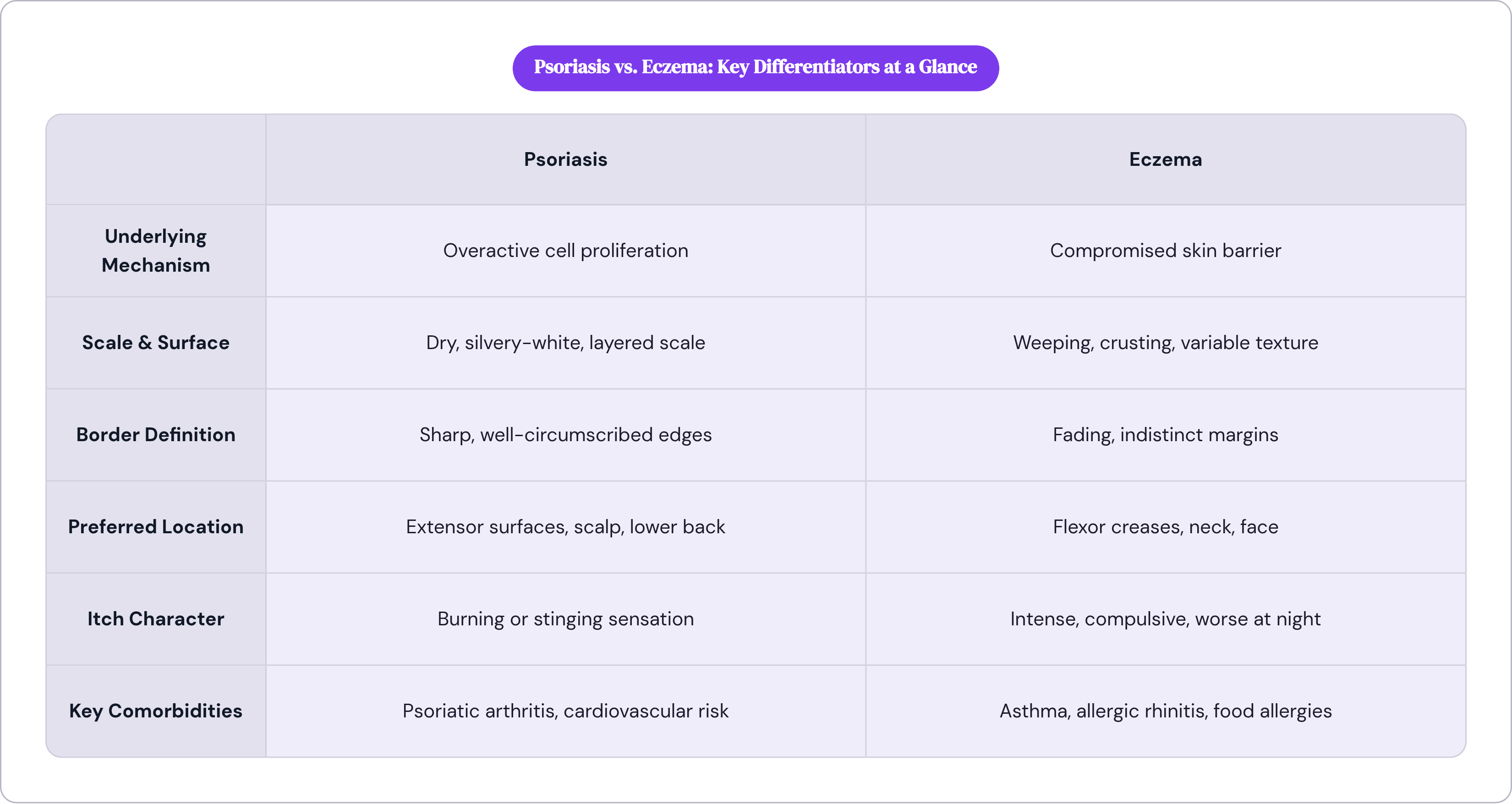
Sharp borders and silver scale point to psoriasis; weeping, fading edges signal eczema.

Start with the biology, because the biology predicts the visual picture, and the visual picture is where almost every misdiagnosis begins.
Psoriasis is a proliferation problem. The immune system, driven by overactive T-cells along a Th17/IL-23 signaling axis, forces skin cells to mature in three to four days instead of the normal twenty-eight to thirty. Cells pile up faster than they can shed. That pile-up becomes a plaque.
Eczema, or atopic dermatitis, is a barrier problem. Mutations in genes like filaggrin compromise the skin's structural integrity, creating microscopic gaps that let allergens and irritants through. The inflammation is the immune system's downstream response to a leaky barrier, not a runaway growth cycle.
Both outputs look like red, itchy, flaking skin at a glance. That surface similarity is where confusion starts and, frankly, where it stays for far too long. A 2024 patient-perspective study documented a 90% initial misdiagnosis rate for psoriasis. That figure isn't a rounding error; it's what happens when you read the surface without reading what's underneath it.
Proliferation creates thick, organized, structured scale. Barrier breakdown creates weeping, crusting, and indistinct edges. Once you see those mechanisms clearly, the visual differences stop looking arbitrary.
The clearest single marker: scale texture and plaque definition
Psoriasis has edges. Eczema doesn't. That single observation does more diagnostic work than almost anything else, and I find clinicians still underuse it.
Psoriatic plaques are raised and thickened, covered in dry, silvery-white scale that looks almost laminated, layered in a way that catches light differently than normal skin. The borders are sharp. Clinicians call them well-circumscribed, meaning you could trace the edge of a lesion with a pen and not lose the line. That precision directly reflects the organized, proliferative mechanism underneath.
Eczema patches look wetter and more variable. The surface weeps or crusts during a flare. Borders fade at the margins rather than stopping cleanly, a direct visual expression of diffuse inflammation spreading through compromised skin rather than cells stacking in a defined zone.
Here's where it gets complicated, though. Chronic eczema that has been scratched repeatedly over months or years can thicken into a leathery texture called lichenification. It can superficially resemble psoriasis, and I've seen confident misreads made on exactly this presentation. But the history usually clarifies things. Visible excoriation marks, scratch lines on the skin surface, and a patient's description of an itch that simply would not leave them alone reframe the picture quickly.
Dry, silver-white flakes with a mappable edge: psoriasis. Weeping, crusted, fading margins: eczema. That shorthand isn't perfect, but it gets you most of the way there before you've asked a single question.
Where on the body each condition shows up — and where they overlap
Location is nearly as diagnostic as texture. It follows logically from the mechanism, which means it's learnable, not arbitrary.
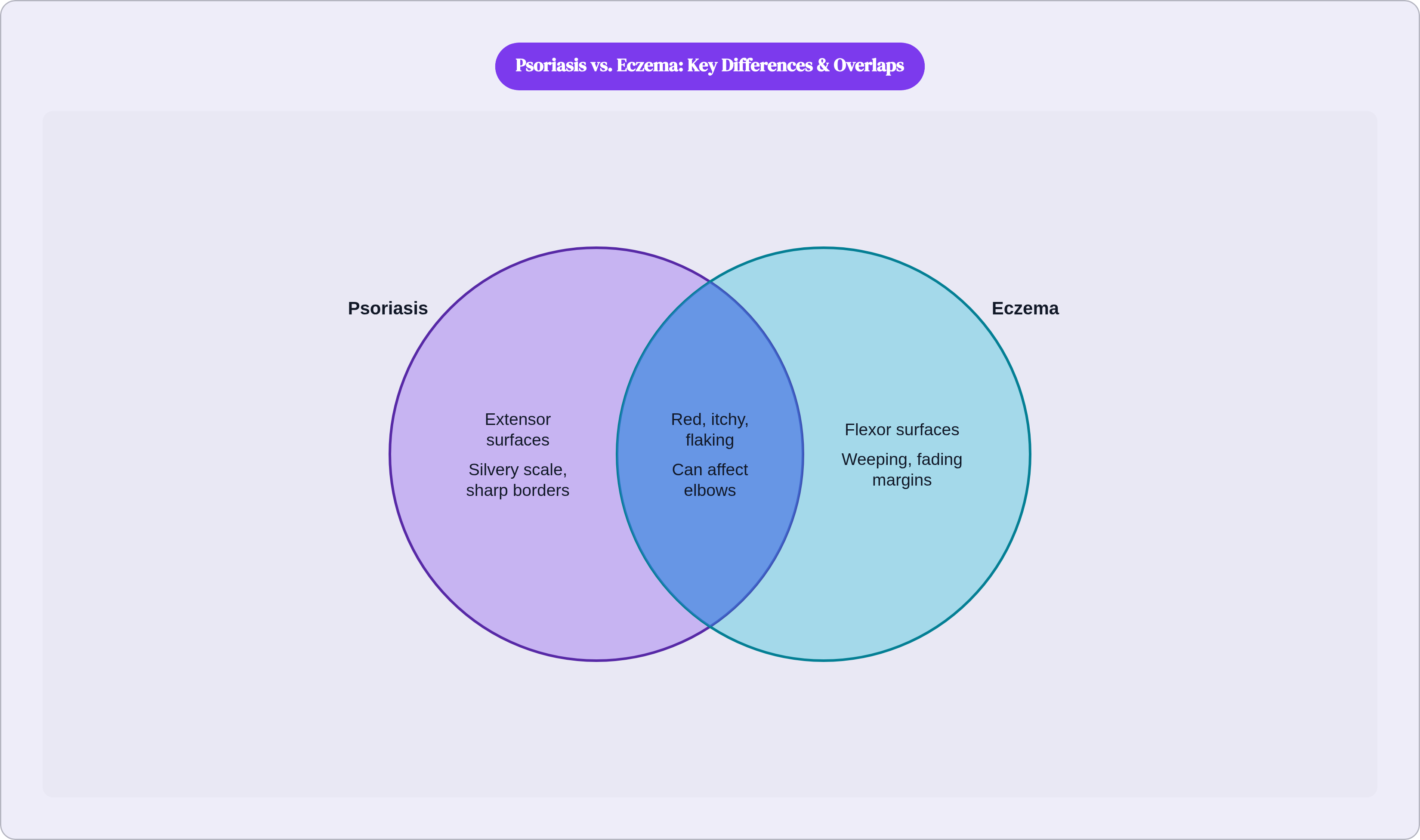
Psoriasis favors extensor surfaces: the outside of the elbows, the outside of the knees, the scalp, the lower back. Surfaces that face outward and bear friction. Eczema favors flexor surfaces: the inside of the elbows, the backs of the knees, the neck, the face. The creases and folds where skin meets skin.
The elbow and knee are the key ambiguity. Both conditions can appear in that general region, and this is where a lot of confident misreads happen. The differentiating detail is specific: psoriasis sits on top of the joint; eczema sits in the crease behind it. Flexural eczema is the clinical term, and patients with it are frequently told their skin "doesn't look like eczema," when it actually looks like textbook eczema in an unfamiliar location.
Scalp involvement skews heavily toward psoriasis. Facial involvement, particularly in infants and young children, skews heavily toward eczema. Neither rule is absolute, but both are reliable enough to factor into the clinical picture.
One signal that gets persistently underweighted: psoriatic arthritis develops in roughly 30% of psoriasis patients. Joint stiffness or swelling alongside extensor surface plaques isn't a separate problem to sort out later in a different appointment. It's meaningful diagnostic information sitting right there in the same conversation.
Itch quality and sensation as a supporting clue
Itch is present in both conditions, but the character of it differs in ways patients can usually articulate if they're asked the right questions. They often aren't.
Eczema itch is intense, frequently described as compulsive and unbearable, worse at night. The itch-scratch cycle isn't a metaphor; it's a visible physical pattern that leaves excoriation marks behind. Sleep disruption affects the vast majority of eczema patients, which is itself a proxy for how relentless the nighttime itch becomes.
Psoriasis itch exists, but is considerably less severe. Patients more often describe it as burning or stinging rather than that irresistible urge to scratch. Sleep disruption in psoriasis is real but meaningfully less prevalent.
Sensation alone cannot diagnose either condition. Both vary considerably between individuals, and itch intensity shifts with flare severity. But when itch quality fits cleanly into one of these patterns, it adds real weight to the overall clinical picture without overriding the visual and structural signals.
How appearance changes on darker skin tones — and why this is routinely missed
Standard clinical descriptions default to lighter skin. Eczema described as pink or red. Psoriasis as red with silver scale. Those descriptors can fail completely on deeper skin tones, and the failure is documented. The National Psoriasis Foundation has found that members of racial and ethnic minority groups are 112% more likely to be living with undiagnosed psoriasis. That number traces directly back to visual misreads at the point of first contact.
On darker skin tones, eczema appears dark brown, gray, or violet rather than red. Psoriasis presents as light-to-dark brown or violet patches, sometimes without any obvious color change at all, showing instead as areas that are thicker, shinier, or differently textured without the classic red-silver contrast. Clinicians mistake these presentations for fungal infections, or miss them entirely because they're reading for the wrong visual cues.
Dyspigmentation compounds the problem. Both conditions leave post-inflammatory color changes, hyperpigmentation or hypopigmentation, that can be more prominent and more persistent on deeper skin tones. That color shift can outlast the active rash, making it difficult to track whether a condition is responding to treatment.
What remains reliable across all skin tones is structure: texture, thickness, border sharpness, scale quality. Color is not the stable anchor it's taught to be. If a clinician's description doesn't match what you're observing on your own skin, naming that discrepancy explicitly during the visit is not an overreach. High-resolution photographs capturing texture under good natural light can bridge the gap between what you see at home and what a provider catches in a brief appointment.
What each condition travels with — the comorbidity picture
Neither condition is only a skin problem, and the direction of systemic risk differs in ways that carry real diagnostic weight.
Psoriasis projects its immune overactivity well beyond the skin. Psoriatic arthritis is the most well-known comorbidity, but established associations also include cardiovascular disease and metabolic syndrome. The same inflammatory signaling driving plaques on the surface is simultaneously driving inflammation elsewhere in the body.
Eczema's systemic pattern runs in a different direction. The condition is strongly associated with asthma, allergic rhinitis, and food allergies, a cluster clinicians call the atopic march, reflecting a shared underlying vulnerability in barrier function and immune regulation that expresses itself across different organ systems over time.
The contrast here is worth sitting with concretely: a patient with joint pain, extensor surface plaques, and a family history of psoriasis looks very different from a patient with childhood asthma, flexural rashes, and seasonal allergies. Those constellations are meaningful clinical data. Treating them as unrelated problems in separate appointments is one of the more consequential ways the systemic picture gets missed for years.
How clinicians confirm a diagnosis when the visual picture is still unclear
A dermatologist reading a rash is synthesizing multiple streams simultaneously: color, thickness, scale, border sharpness, distribution, history, patient-reported symptoms. No single visual feature is diagnostic on its own.
Medical history adds a dimension that visuals alone cannot provide. Psoriasis has a strong genetic component, including associations with specific immune-related gene variants, so family history matters. Known allergies, identifiable triggers, and the pattern of flares over time inform the picture in ways a single clinical photograph cannot.
When the visual presentation is ambiguous, a skin biopsy is the clarifying tool. A small sample examined under a microscope can distinguish psoriatic proliferation from the inflammatory changes characteristic of eczema. It isn't always necessary, but when the picture is murky, it removes the guesswork.
One more complication worth naming plainly: it is possible to have both conditions simultaneously. Some psoriasis treatments trigger a secondary eczematous response, which complicates everything considerably. "Treated but still flaring" is not always treatment failure. Sometimes it's a second condition emerging in response to the first being addressed, and conflating the two sets back the entire diagnostic timeline in ways that are hard to recover from quickly.
Getting a qualified eye on a rash without waiting weeks for an in-person appointment
Teledermatology has changed the practical timeline for getting a qualified assessment. Published evaluations of high-resolution photo analysis combined with structured history intake show diagnostic accuracy for common skin conditions, including eczema and psoriasis, landing in the low-to-mid 90s. When the alternative is a weeks-long wait for a dermatology referral while remaining on the wrong treatment, that accuracy range matters.
Asynchronous consultations, where a patient uploads photos and completes a symptom history and a clinician reviews without a synchronous call, typically return an evaluation within 24 to 48 hours. What the clinician is reading in that review: scale texture, border definition, rash distribution, itch quality, location relative to flexor or extensor surfaces, and comorbidities. The same structural features that differentiate these conditions in person.
Teledermatology cannot perform a biopsy, assess skin thickness by touch, or definitively evaluate complex presentations requiring hands-on examination. When a biopsy is indicated, a responsible telehealth encounter says so plainly. Patients should know where that boundary sits before they arrive at it.
Photo quality is not incidental. Capturing texture and border sharpness in good natural light, from multiple angles, gives a reviewing clinician the best possible chance of reading the structural signals that actually differentiate these conditions. A blurry photograph in dim light is barely better than a verbal description of something you're struggling to articulate.
The structured symptom intake matters equally. Knowing whether a rash sits on an extensor or flexor surface, whether the itch is compulsive or burning, whether the borders are mappable or diffuse, whether there's joint involvement or a history of asthma: these details transform a vague complaint about "a rash" into something a clinician can actually work with. Getting specific about structure before you try to describe something is, in my experience, the difference between an appointment that moves and one that doesn't.