Identifying Fungal Skin Infections by Appearance
Two broad categories of fungi create distinctly different visual patterns on skin.

Two broad categories of organisms cause most fungal skin infections, and the difference between them changes everything you see on the skin surface.
Dermatophytes are molds, specifically genera like Trichophyton, Microsporum, and Epidermophyton. They eat keratin, the structural protein composing the outermost layer of skin, hair, and nails. Most dermatophytes stay confined to that stratum corneum, which is why their signs are surface phenomena: scaling, color shifts, active borders. The damage is architectural. Yeasts are a different story. Candida and Malassezia furfur both live on most people's skin without causing any particular problem until something tips the local environment in their favor. Warmth. Moisture. A disrupted skin barrier. Immune suppression. They're not foreign invaders the way a dermatophyte is; they're opportunists waiting for conditions to shift.
That distinction, dermatophyte versus yeast, shapes not just treatment but what you're actually looking at.
Location is your first clue, and it's the one most people skip. Fungi colonize specific microenvironments: skin folds with sustained friction and moisture, keratinized surfaces like nails and scalp, sebum-rich zones across the trunk. Before you assess shape or color, ask where exactly this rash is sitting. That answer alone narrows the differential considerably. Trichophyton rubrum dominates adult infections broadly; T. tonsurans leads in pediatric scalp and body presentations. Even pathogen prevalence is age-stratified, which tells you something about how precisely adapted these organisms are to their hosts.
Ringworm on the body: the ring shape and what's happening at the border
Tinea corporis has a presentation almost designed to be memorable, but the name can mislead you into thinking any ring-shaped rash is ringworm. The shape matters less than what's happening at the edge.
The fungus spreads outward from a central point. As it moves, the immune system catches up with the center and partially clears it. What you're left with is an annular lesion: a circular or oval patch with a raised, red, scaly outer border and a relatively clear center. That border is where the fungus is actively colonizing, where the scale is densest and the redness most concentrated. Lesions typically range from one to five centimeters, though multiple rings can merge into larger, irregular patches, especially in children who've picked up the infection from a pet or a classmate.
The centrifugal activity pattern is the feature worth internalizing. Not just that the rash is round, but that the edge is more inflamed than the middle.
The mimics are worth knowing cold. Eczema is itchier, less geometrically organized, and lacks that raised ring structure. Annular psoriasis shares the ring shape but comes with thicker, more silvery scale and a different distribution. Pityriasis rosea usually starts with a single larger herald patch before spreading in a Christmas-tree pattern down the back. And secondary syphilis, a clinically important entry on this list, can produce annular lesions convincingly enough that an unusual or persistent ring-shaped rash warrants professional assessment rather than a pharmacy run.
If someone has applied a steroid cream to a ring-shaped rash and it's spreading rather than resolving, that behavior alone should shift suspicion toward fungal infection.
Athlete's foot: three distinct patterns that don't all look alike
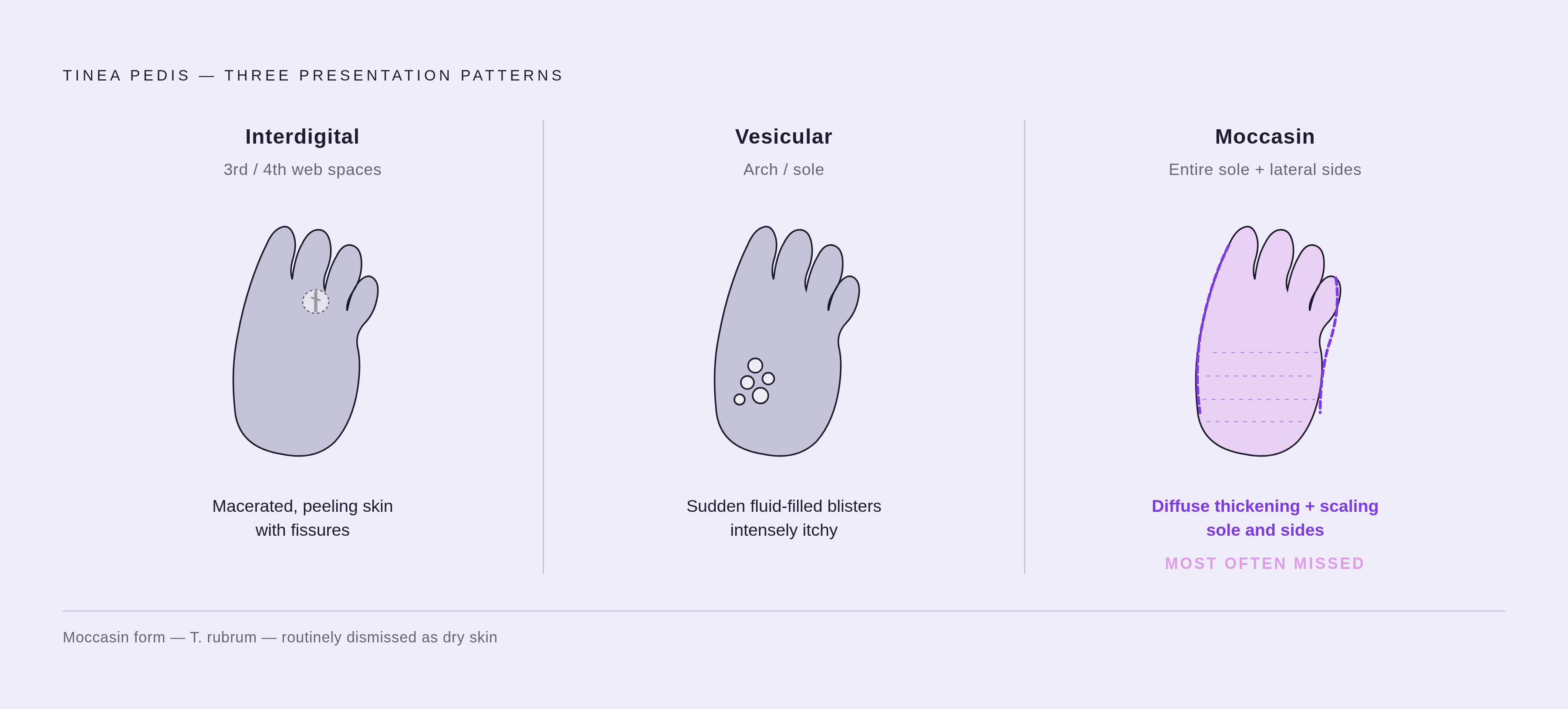
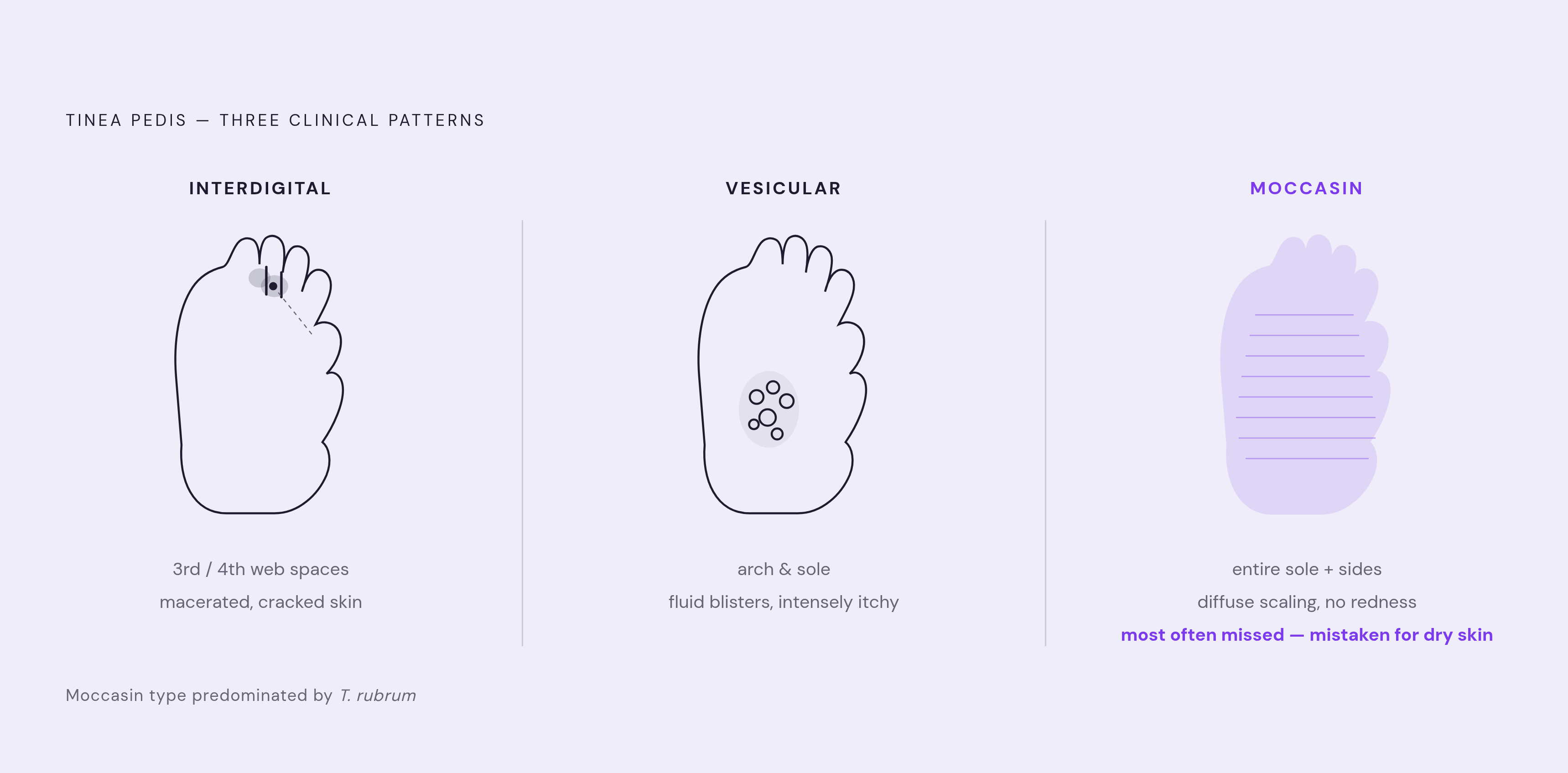
"Athlete's foot" implies a single, recognizable presentation. The reality is that tinea pedis presents in three visually distinct patterns, and conflating them means missing the diagnosis on at least two of them.
The interdigital form is the most common. It lives between the toes, particularly the third and fourth web spaces. The skin appears whitened, softened, macerated, with peeling and cracking. In acute flares, painful fissures or small blisters show up. Redness can be vivid, but the dominant feature is that waterlogged, broken-down skin texture.
The vesicular form is the most dramatic: a sudden eruption of fluid-filled blisters on the arch or sole, intensely itchy, acute in onset. This one can look like contact dermatitis or an id reaction, an immune response to a fungal infection elsewhere on the body, which makes it easy to chase the wrong diagnosis entirely.
The moccasin form is the one most reliably missed. No acute redness, no blistering. The skin across the entire sole thickens and scales diffusely, wrapping up the sides of the foot in the distribution of a low-cut moccasin shoe. T. rubrum is the predominant pathogen here. Many people dismiss it as dry skin and do exactly that for months or years, until it progresses into something that can't be ignored. Untreated, tinea pedis can lead to secondary bacterial infections including cellulitis; in vulnerable populations, the consequences escalate further.
One more variable that clinical literature underemphasizes: skin tone modifies what you see. Depending on baseline pigmentation and severity, affected areas appear red, white, gray, or purplish. Redness is not a universal sign, and treating it as one produces missed diagnoses.
Jock itch and scalp ringworm: how age and anatomy shape what you see
Two more dermatophyte infections, both familiar by name, both with presentations shaped heavily by anatomy and the age of who they affect.
Tinea cruris favors the inner thighs and groin folds, sometimes extending to the buttocks. Like tinea corporis, it presents with a raised, scaly, well-demarcated border, but because it follows the groin fold, its shape runs toward a crescent or half-moon rather than a full circle. A useful distinguishing detail: jock itch characteristically spares the scrotum. Candidal intertrigo in the same region involves the scrotum more fully and looks more uniformly red and moist. That anatomical specificity is a practical sorting variable. Tinea cruris is predominantly a condition of adolescent and adult males, uncommon before puberty.
Tinea capitis is essentially the demographic mirror image. Almost exclusively a disease of prepubertal children; something about the hormonal shifts at puberty, likely changes in sebum composition, appears to confer resistance in adults. Scalp presentations vary more than most people expect. Some cases show scaly, inflamed patches with hair loss within the patch. Others present with infected hair shafts that break off at the scalp surface, leaving a "black dot" pattern. The most severe form, a kerion, presents as a boggy, pustular, painful mass that looks almost like an abscess and can cause permanent scarring hair loss if treated promptly.
The critical mimic here is alopecia areata, which also produces patchy hair loss. Alopecia areata is smooth, without scaling or significant inflammation. The treatments for the two conditions aren't just different; they are physiologically opposed. Treating tinea capitis with immunosuppressants, which alopecia areata requires, worsens the infection. Getting this one right matters in a concrete, immediate way.
Tinea versicolor: the infection that changes color with the season
Tinea versicolor operates on a completely different logic from the ring shapes and active borders of dermatophyte infections. Its whole presentation is organized around pigment disruption. The causative organism, Malassezia furfur, is a yeast native to skin that overgrows in hot, humid conditions. Its preferred terrain is the sebum-rich upper trunk: back, chest, shoulders, upper arms, neck.
What it produces visually looks nothing like ringworm. No ring, no raised border. Instead, discrete patches, sometimes numerous and sometimes confluent, that are lighter or darker than surrounding skin. White, tan, pink, brown, depending on the patient's natural skin tone and recent sun exposure.
The seasonal behavior is one of its most useful identifiers. In summer, when surrounding skin tans, the affected patches don't. They fail to darken and stand out as conspicuously pale. In winter, the same patches appear comparatively darker against a lightened baseline. The rash literally shifts its visual relationship to surrounding skin based on season and sun exposure. Once you've seen it work that way, you don't forget it.
Itch is mild or absent. Many people notice the color change long before they notice any discomfort, which is part of why it goes unaddressed for so long. Look closely or lightly scrape the surface, and a fine, powdery scale becomes visible. Subtle, but consistent.
The common misread is vitiligo, which also causes hypopigmented patches. Vitiligo involves complete pigment loss, characteristically sharp edges, and no scale at all. Tinea versicolor patches have softer edge definition and carry that subtle scale. Learnable, though not always obvious on first glance.
Cutaneous candidiasis: what skin-fold infections look like and who gets them
Candida lives on most people's skin without incident until something shifts: sustained heat and moisture, a compromised skin barrier, immune suppression, persistently elevated blood glucose. The locations it favors follow moisture gradients directly. Armpits, under the breasts, abdominal skin folds, the groin, the diaper area in infants. Anywhere skin presses against skin and traps humidity.
The rash is typically bright red, moist, sometimes raw-looking. The center of the fold can appear macerated, whitened, almost waterlogged. But the feature that most reliably distinguishes candidal intertrigo from a simple irritant rash is the presence of satellite lesions: small red spots or pustules scattered just outside the main rash border, as if the infection is sending out isolated advance positions. When you see those satellites, Candida moves to the top of the differential.
Diabetes amplifies risk significantly. Research suggests that up to 70% of diabetic patients develop skin lesions related to deterioration in skin barrier function and changes in skin pH; candidal infections are among the most common fungal complications in this population. Obesity increases susceptibility for a related reason: deeper and more numerous skin folds create more territory.
In men, Candida can manifest as balanitis, redness, white patches, and soreness under the foreskin. In women, vulvovaginal candidiasis presents with intense itch and curd-like discharge. These aren't skin-fold infections in the strict anatomical sense, but the underlying biology is continuous with cutaneous candidiasis.
Nail fungus: the visual signs that separate it from ordinary nail damage
Onychomycosis affects toenails far more often than fingernails, and the mechanics make immediate sense: toenails spend hours each day in enclosed, warm, moist environments.
The classic visual progression begins at the distal edge, the tip or sides, and moves toward the base. The nail thickens. It discolors to yellow, white, brown, or a chalky, opaque tone. Edges become brittle and crumble. In some cases the nail plate separates from the nail bed, a condition called onycholysis, leaving a lifted, opaque section that's uncomfortable and a potential entry point for secondary infection.
Here's the complication: trauma-related nail dystrophy looks nearly identical. Runners, people in ill-fitting shoes, anyone subjecting their toenails to repetitive low-grade impact can develop thickened, discolored, crumbly nails that are clinically indistinguishable from onychomycosis by appearance alone. The American Academy of Family Physicians has noted explicitly that visual assessment is insufficient to differentiate the two reliably, which is why confirming with a KOH preparation or culture is specifically recommended before initiating systemic antifungal therapy. Systemic antifungals carry real side-effect profiles. Prescribing them without confirmation, when the underlying condition is mechanical rather than infectious, is a clinically indefensible approach.
Prevalence of onychomycosis in diabetic patients reaches approximately 29%, and that population faces meaningfully elevated overall risk of mycotic infections compared to the general population.
Visual features that cut across all fungal infections and help distinguish them from look-alikes
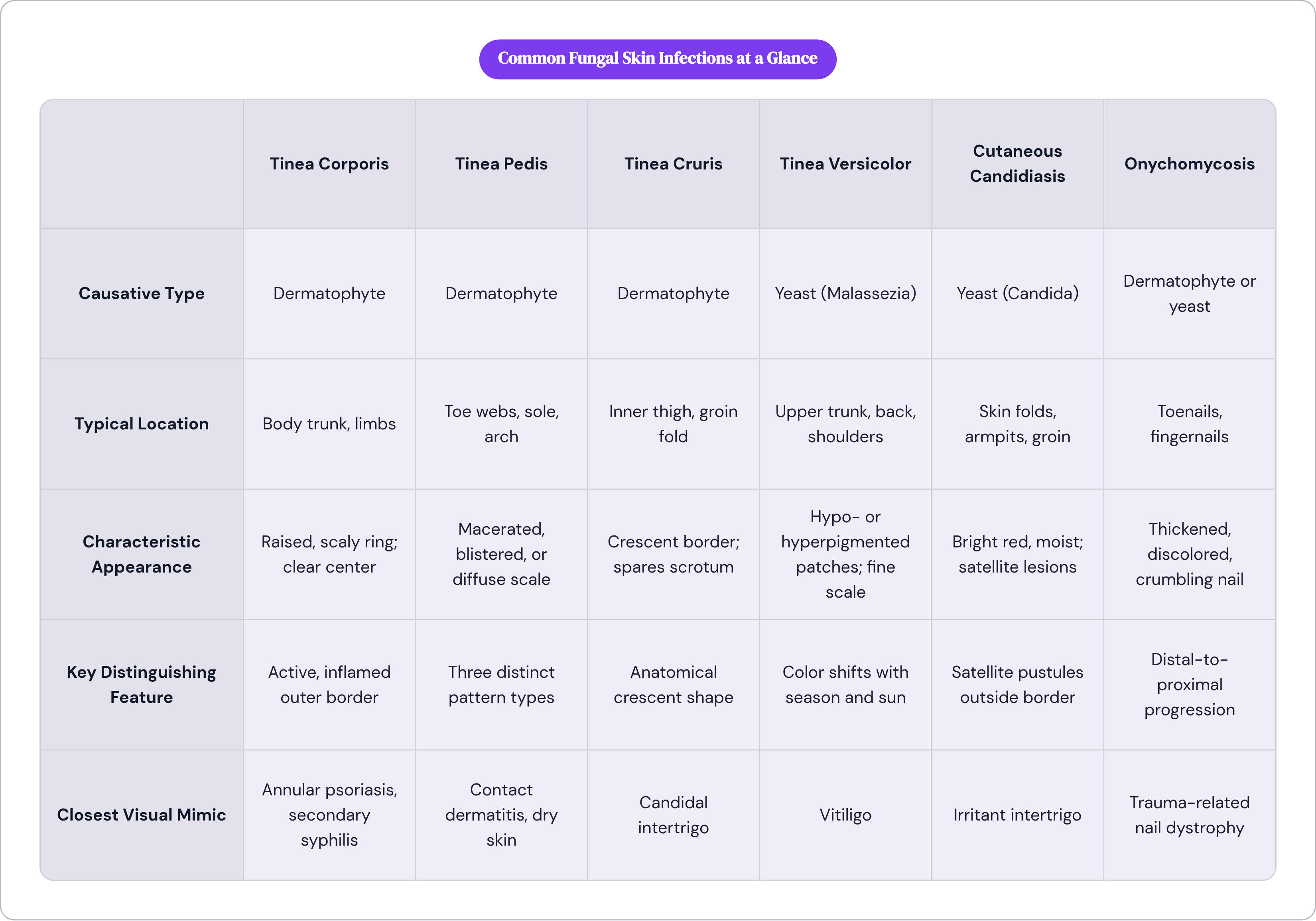
Step back from the individual presentations and a few consistent principles hold across all of them.
Location comes first. The anatomical zone narrows your list immediately, and each zone has a short, predictable list of candidates. Color and shape assessed without location context carry much less diagnostic weight than most people assume.
Border behavior separates dermatophytes from Candida in most cases. An active, raised, scaly border that is more inflamed than the center suggests a dermatophyte expanding outward. Diffuse, moist redness without a clear border, typically in a fold, suggests Candida.
Scale texture carries its own diagnostic signal. Tinea corporis and tinea pedis produce dry, flaky scale. Tinea versicolor produces a fine, almost powdery scale that requires close inspection. Candidal infections produce little or no scale; the surface is often macerated instead.
Satellite lesions are Candida's specific calling card. Small pustules or spots outside the main rash perimeter point toward Candida in a way that no dermatophyte infection reliably mimics.
Pigment change without significant inflammation is tinea versicolor's signature. If the dominant complaint is color rather than itch, and the location is the upper trunk, that two-variable combination is meaningful.
What fungal infections generally don't do is equally worth knowing. They rarely produce the weeping, honey-colored crusting of bacterial infection. They also leave the hard, raised, silvery-scaled plaques of psoriasis alone. And when a rash that presents like eczema keeps spreading or worsening under steroid treatment, that behavioral signal is one of the most practically useful clues available.
One variable deserves direct acknowledgment: on darker skin, the redness of fungal infections presents as brown, gray, or purple rather than the classic red described in most clinical literature. Color as a single indicator is less reliable across diverse skin tones than location, border shape, and texture assessed together.
When a visual assessment needs clinical confirmation
Even trained clinicians misidentify fungal infections with regularity. The mimics are numerous, the visual overlap is genuine, and the conditions that modify appearance, skin tone, immune status, concurrent treatment, are in play more often than not.
A prospective cohort study from Singapore found that microscopy identified 25.0% of skin scrape samples as positive, while fungal culture identified 16.7%; when both methods were used together, concordance reached 84.5%. Even laboratory methods used in isolation miss a meaningful proportion of cases. Visual assessment alone sits somewhere below that.
For nail fungus and scalp ringworm specifically, clinical guidelines recommend laboratory confirmation before initiating systemic treatment. These are situations where treating empirically on appearance carries real risk: the wrong drug, unnecessary side effects, continued progression of an infection being addressed for the wrong organism.
Some presentations warrant professional assessment regardless of how confident the visual read feels. A rash on the scalp, nails, or face. An infection spreading despite a reasonable trial of over-the-counter antifungal. Signs pointing toward a more serious mimic, particularly secondary syphilis. Any fungal infection in someone with diabetes, compromised immunity, or poor peripheral circulation.
For straightforward presentations, a classic ring on the forearm or the typical interdigital softening of athlete's foot, a precise visual description communicated to a clinician can meaningfully shorten the path to correct treatment. Location, border behavior, texture, scale quality, and how the rash has changed over time. Described accurately, those details are more useful to a clinician than "I have a rash and it itches."