How Skin Conditions Present Differently on Dark Skin

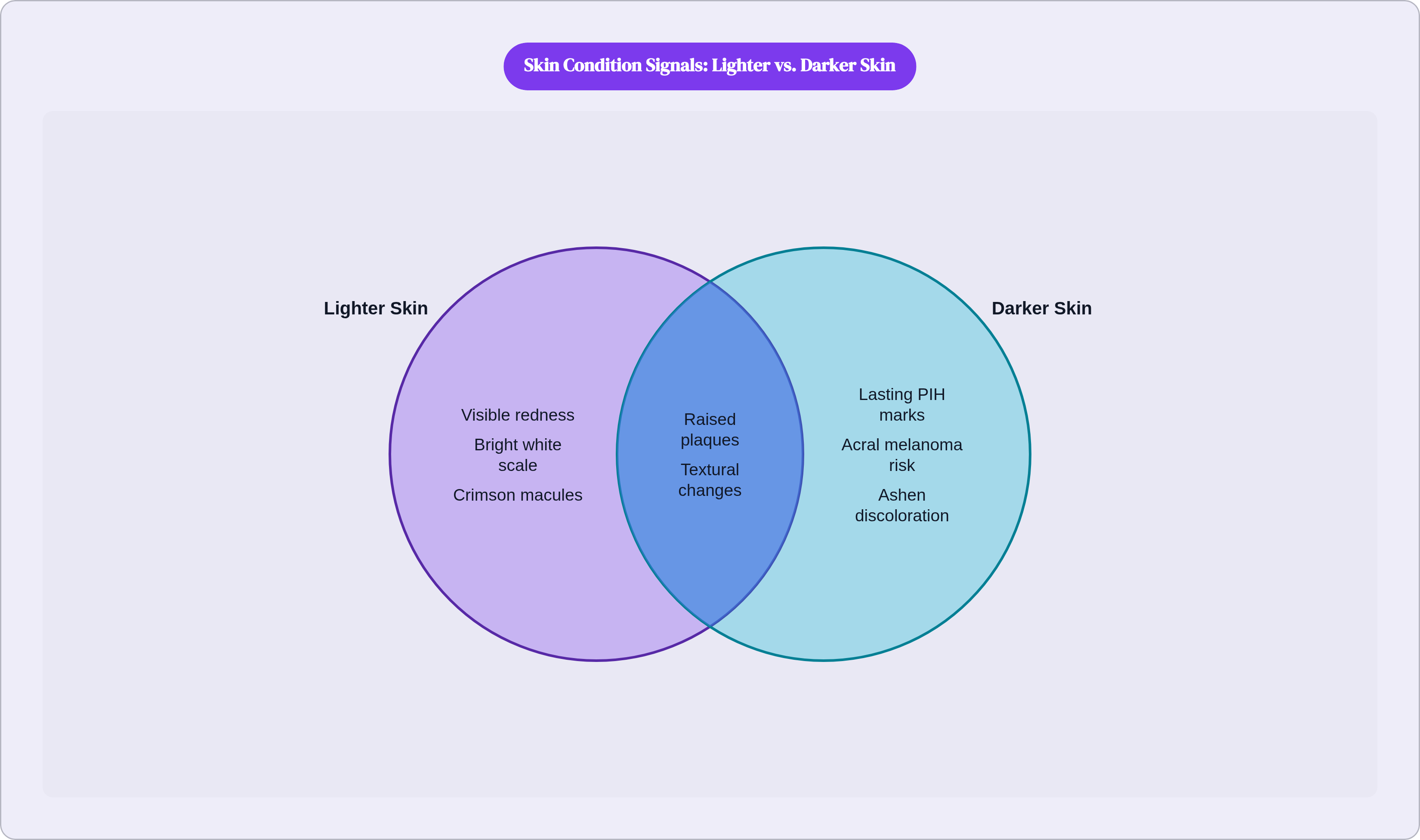
Open almost any dermatology textbook. The eczema image shows a red, weeping patch on fair skin. Psoriasis is a vivid red plaque with bright white scale. A drug rash is a field of crimson macules spreading across a pale trunk. These images are not wrong. They accurately represent what those conditions look like on the skin they were photographed on. The problem is that they were never meant to be universal, and somewhere along the way, they became treated as if they were.
Studies of dermatology teaching materials have found, consistently, that images of darker skin make up only a small fraction of the total. Single digits, in many cases. The result is a training pipeline, running from medical school through residency, in which clinicians see thousands of images on lighter skin and comparatively few on darker skin. Many clinicians will tell you this plainly when asked: they weren't trained to recognize what they weren't shown.
For patients, the consequence is immediate and disorienting. When you search your symptoms or compare your skin to a clinic pamphlet, the image doesn't match what you're seeing. If the defining visual marker is redness and your skin doesn't show redness the way the photograph does, you conclude you don't have the condition. Or you feel confused. Or you walk out of an appointment wondering whether something is wrong with your ability to read your own body.
Here is the biology, because it changes everything. Melanin, the pigment that determines skin tone, is present in higher concentrations and more reactive forms in darker skin. When inflammation occurs, the underlying redness is generated by increased blood flow beneath the surface. On lighter skin, that signal travels through relatively little pigment and becomes visible as redness. On darker skin, the melanin layer is thick enough to absorb or mask that red wavelength. What shows through instead is the effect of inflammation on the pigment itself: shifts toward purple, gray, brown, or ashen tones. Textural changes. Swelling. And often lasting pigmentary aftermath that outlasts the original condition by months.
This is not an edge case. It is the default experience for a large and growing share of the population. Color, and redness specifically, is an unreliable guide. What to watch for goes well beyond "is it red?"
The rest of this piece works through that, condition by condition.
What eczema actually looks like on darker skin
If you have darker skin and you have eczema, there is a reasonable chance you didn't recognize it the first time. The textbook image, that red weeping patch, doesn't describe what most people with darker skin experience.
The inflammation presents as brown, purple, gray, or ashen discoloration rather than redness. In some presentations, the area won't look dramatically different in color from the surrounding skin at all. What distinguishes it is everything else.
Persistent itching is usually the most reliable signal, often intense and frequently worse at night. The rash appears as small raised bumps in a papular or follicular pattern, not the flat inflamed patch shown in most educational materials. Skin thickening, lichenification in clinical terms, develops because the itch-scratch cycle damages the skin barrier repeatedly. Texture changes, roughness, scaling, dryness, are often more diagnostically useful than color.
Around the eyes, eczema can produce scaly, hyperpigmented plaques on both upper and lower lids. This presentation is more common and more prominent in darker-skinned patients, and it is frequently mistaken for contact dermatitis or allergic reaction. Persistent scaling or darkening around the eye area without an obvious cause puts eczema on the list.
Then there's the aftermath. Post-inflammatory hyperpigmentation can be as distressing as the eczema itself. Dark marks following a flare can persist for months after the skin has healed, a visible record of each episode. Early recognition and treatment matter precisely because they reduce the cumulative pigmentary damage over time.
The absence of redness is not the absence of eczema. Persistently itchy, rough, thickened skin producing small raised bumps that don't resolve warrants attention regardless of whether anything looks red.
What psoriasis looks like on darker skin — and why it's frequently mistaken for other conditions
Psoriasis on lighter skin has a recognizable signature: raised plaques, vivid redness, white or silvery scale. On darker skin, that signature changes substantially. The plaques are still raised and thickened, but the color shifts to purple, brown, or gray. The scale appears silver or gray rather than the bright white that photographs so distinctly on fair skin. The overall picture can look nothing like anything labeled "psoriasis" in a standard reference.
This creates a genuine diagnostic problem. Psoriasis on darker skin shares visual features with eczema, lupus, lichen planus, and fungal infection, and without redness to orient the differential, distinguishing between them requires more careful examination, a detailed history, and sometimes a biopsy. Misdiagnosis is common. It carries real costs: delayed treatment, inappropriate therapy, extended pigmentary damage from uncontrolled inflammation.
There is an additional subtlety worth understanding. On darker skin, the visible signs of active psoriasis persist longer after the underlying inflammation has actually resolved. The skin holds a pigmentary memory of the episode even when the pathology has quieted. A patient who concludes that treatment isn't working when it is operating on this dynamic is drawing from a misleading signal.
That same persistence has a practical implication: treating early and consistently, before plaques have time to deepen, leaves a smaller pigmentary footprint.
Watch for thickened, raised skin with any silvery or gray scale; patches that persist and don't resolve over weeks; itching or discomfort even without obvious redness. Those features together warrant evaluation.
How rosacea and drug rashes show up when redness doesn't
Rosacea is defined, in most clinical frameworks, partly by visible redness and flushing. That dependence on redness as a diagnostic criterion creates a structural problem for darker-skinned patients: the primary visual signal is harder to detect and, in some presentations, functionally invisible.
On darker skin, rosacea-associated inflammation manifests as subtle tonal shifts, warmth to the touch, burning or stinging, and textural change rather than vivid flushing. The condition goes unrecognized for years, attributed to other causes, because clinician and patient alike are looking for something that isn't showing up in the expected form. Papulopustular rosacea, the subtype characterized by bumps and pustules, will be more readily identified because the textural component is apparent; the erythematous component, though, will still be muted.
Drug eruptions tell a similar story. Maculopapular rashes from medication reactions present on lighter skin as vivid red macules spreading from the trunk outward. It's a visually striking pattern, taught as a classic clinical sign. On darker skin, those same eruptions appear purple or gray, are significantly less vivid, and are difficult to distinguish against surrounding skin. They are frequently underappreciated in clinical settings as a result.
The useful principle for both conditions: report change. An area that feels warmer than surrounding skin. A texture that has shifted. A sense of burning or flushing without an obvious explanation. Clinicians are better oriented by a precise description of what has changed than by the absence of an expected visual finding.
Cutaneous lupus on darker skin — a presentation often centered on pigment rather than color
A substantial majority of people with lupus develop skin symptoms at some point, which makes skin presentation a front-line diagnostic clue for what is ultimately a systemic condition. Getting the skin manifestations right matters well beyond dermatology.
On darker skin, lupus rashes appear as dark purple or dark brown rather than the bright red described in most textbooks. The classic butterfly rash across the cheeks and nose, a vivid erythematous finding on lighter skin, will be subtle, hyperpigmented, or nearly undiscernible by color alone.
Discoid lupus erythematosus, DLE, is particularly important here. It is more prevalent in Black women and follows a more severe course in that population. Lesions concentrate on sun-exposed areas, with the scalp and ears being especially common sites. Within those lesions, depigmentation is frequent, producing a distinctive pattern: darkened borders with lighter or scarred centers. Lesions are typically annular, ring-shaped, with post-inflammatory hyperpigmentation common at the margins.
Scarring alopecia is the complication that gives DLE its particular urgency. Once follicles are destroyed by inflammation, hair loss is permanent. This makes early diagnosis, before significant scarring has occurred, consequential in a way that goes beyond appearance.
Dark purple or brown patches on the face, ears, or scalp, particularly those that scar, leave lasting discoloration, or are accompanied by unexplained hair loss at the hairline, warrant prompt evaluation.
Melanoma in darker skin — where it appears, what it looks like, and why it's caught late
The lower overall incidence of melanoma in darker-skinned populations is real. It has also generated a damaging misconception: that darker-skinned people don't need to monitor for skin cancer. Incidence data is not a reason for complacency. It is a reason for precision.
Survival outcomes for Black patients diagnosed with melanoma are significantly worse than for white patients. That gap is driven primarily by late-stage diagnosis, not by any intrinsic difference in the biology of the cancer. It is a detection problem, and it is downstream of the misconception that darker skin is not at meaningful risk.
The most relevant subtype is acral lentiginous melanoma, ALM. It is uncommon in the general population but is the dominant melanoma subtype among people of Asian and African descent, occurring at rates many times higher than in lighter-skinned groups. The single most important thing to understand about ALM is that it is not related to sun exposure. Standard sun-safety messaging, and standard self-exam guidance focused on sun-exposed areas, misses it entirely.
ALM occurs on the palms, the soles of the feet, and under the nails. On the sole, it appears as a small dark spot, something that resembles dried blood or a scab, or a non-healing sore on dry or cracked skin. It is routinely mistaken for a blood blister, a heel fissure, a callus, a wart. What distinguishes it is persistence: a blood blister resolves. A spot that stays for weeks, or grows, warrants evaluation.
Under the nail, ALM presents as a dark streak running lengthwise along the nail, a finding called longitudinal melanonychia. A benign streak is possible, particularly in darker-skinned people, where it is a more common normal variant. The concerning feature is the Hutchinson sign: pigmentation spreading from the streak onto the surrounding skin of the nail fold. That extension beyond the nail boundary is the warning. A subungual bruise from a known injury will typically resolve as the nail grows; a streak with no associated injury, or one involving the surrounding skin, warrants dermatologic evaluation.
Squamous cell carcinoma in darker skin also follows a different anatomic pattern. Rather than arising on sun-damaged facial skin as it does in lighter-skinned patients, SCC in darker skin more commonly arises on the legs, anogenital regions, and sites of chronic scarring or irritation.
For darker-skinned patients, skin cancer monitoring must include the soles, palms, nails, and areas of chronic skin irritation. Sun exposure is not the relevant variable for the cancers most likely to affect you.
Post-inflammatory hyperpigmentation — the aftermath that often outlasts the condition itself
I want to be direct about something: post-inflammatory hyperpigmentation is not a cosmetic footnote. Clinicians often treat it as one, which is its own kind of diagnostic failure.
PIH is a predictable biological consequence of skin inflammation, and it is more prominent and more persistent in darker skin because of how melanocytes respond to injury. When skin is inflamed or damaged, melanocytes increase their output. On darker skin, those cells are more reactive and produce more melanin in response to the same stimulus. The resulting dark mark can persist for months to years after the underlying condition has resolved. Eczema, acne, psoriasis, lupus, a drug reaction, even a minor abrasion: each can leave PIH behind. The condition heals. The mark stays.
The marks serve as a visible record of each episode, sometimes more conspicuous than the active disease ever was. That visibility is a common driver of treatment-seeking even when the underlying condition is currently quiet, which tells you something about their weight in daily life.
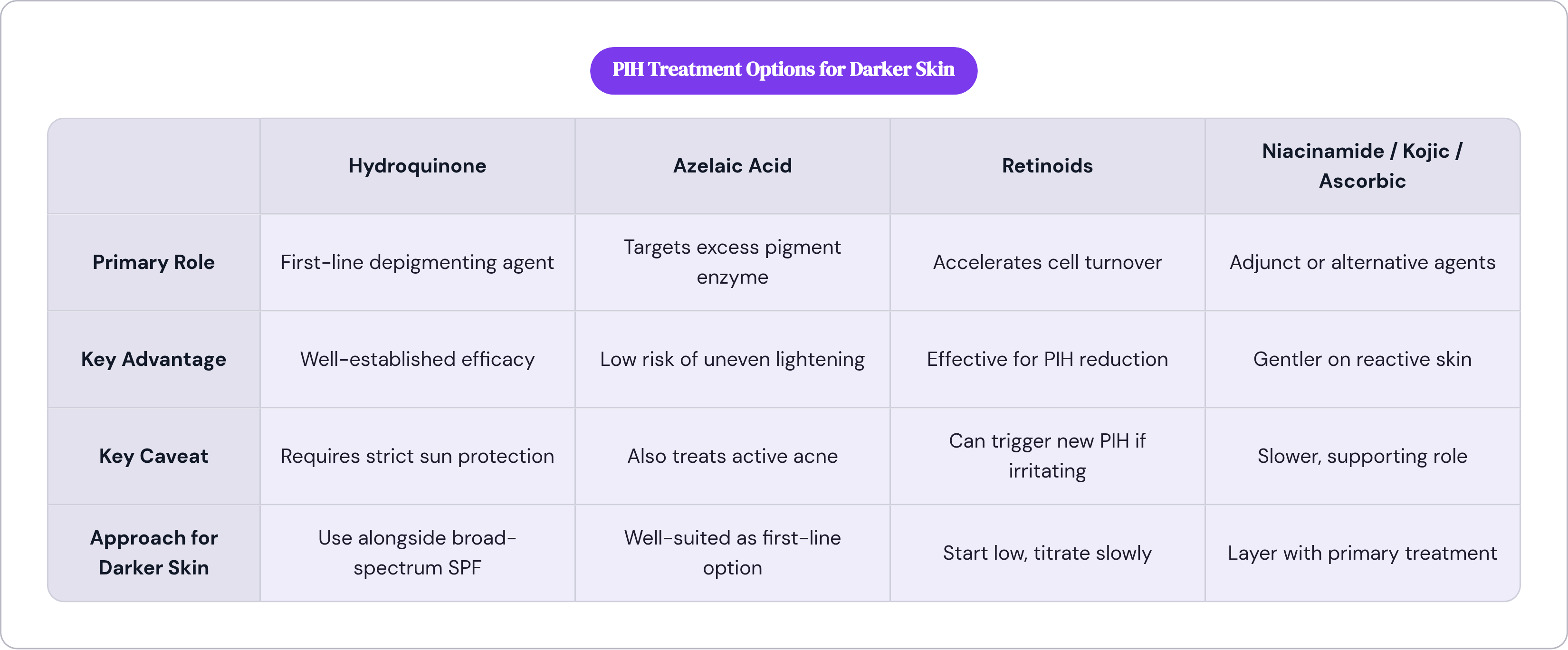
The treatment landscape has real options. Hydroquinone is the most established depigmenting agent and is frequently used as a first-line option alongside rigorous sun protection. Azelaic acid is particularly well-suited to darker skin because it targets the specific enzyme involved in excess pigment production rather than broadly lightening surrounding tissue, which reduces the risk of uneven results; it also addresses active acne simultaneously, a meaningful advantage when PIH and acne coexist. Niacinamide, kojic acid, ascorbic acid, and licorice extract serve as adjuncts or alternatives. Retinoids are effective but carry a relevant caveat: they cause enough irritation to trigger new PIH in darker skin, so starting at low concentrations and titrating slowly is practical necessity, not optional caution.
Sunscreen is foundational. UV exposure deepens existing hyperpigmentation by stimulating further melanin production, so daily broad-spectrum protection is the non-negotiable base of any PIH regimen. Without it, other interventions are working against a headwind.
Improvement takes months of consistent treatment. Patients who understand that timeline are far more likely to stay with a regimen that is working. Abandoning an effective treatment at three weeks because nothing looks different yet is one of the most common ways PIH management fails.
How to describe what you're seeing when you seek care — and what to ask
Most clinical language around skin conditions defaults to redness as the primary visual marker. When you have darker skin, that default leaves you without accurate language for what you're actually observing, and without language it becomes harder to communicate clearly, advocate for yourself, or convey urgency when urgency is warranted.
Specific language closes that gap. Instead of "it's red," try: "it looks purple compared to the surrounding skin," or "the area looks gray or ashen," or "there's a dark patch that appeared after a rash I had a few months ago." Each of those descriptions gives a clinician more to work with than a color term that doesn't match what you're seeing.
Beyond color, describe texture and sensation. Is the skin thickened, rough, raised? Is it itchy, painful, or numb? Has the texture changed even when the color hasn't? Has a mark persisted longer than a normal bruise or irritation would explain? These dimensions carry as much diagnostic weight as color, sometimes more.
For skin cancer concerns specifically, the most important shift is anatomic. Check the soles of your feet, your palms, your nails. Look for dark streaks running lengthwise. Check areas of chronic skin irritation or scarring. These are the locations where the cancers most relevant to darker skin arise, and they are rarely covered in standard skin cancer education materials.
Photographs taken in good natural light capture texture, subtle tonal shifts, and the relationship between a lesion and its borders in ways that verbal description often cannot. They're particularly useful in telehealth contexts. On that note: Nolla is an AI-guided skincare telehealth service where a licensed US clinician reviews your symptoms and can issue a prescription in as little as 10 minutes, rather than leaving you with raw algorithm output. It can help bridge the gap between "something is off" and "I have a specific, documented concern to bring to an in-person specialist." Any finding that warrants concern should still prompt follow-up with an in-person dermatologist.
It is entirely reasonable, when choosing a dermatologist, to ask directly: "Are you comfortable diagnosing and treating skin conditions in patients with darker skin?" That question is not impolite. It is an efficient way to find out whether the person you're about to trust has had meaningful training in what your skin actually looks like.
What the AI tools used in dermatology currently get right and wrong for darker skin
AI-assisted dermatology tools are a real part of the landscape now, and they deserve clear thinking rather than reflexive dismissal or uncritical enthusiasm.
The core problem is the same one running through this entire piece: training data. Machine learning models learn to recognize patterns from the images they were trained on. If those training datasets underrepresent darker skin tones, the model learns to recognize conditions as they present on lighter skin and performs less reliably elsewhere. This pattern has been documented across multiple types of medical imaging AI. Dermatology is not exempt.
Vendors often cite accuracy figures from internal validation studies, and those numbers tend to look reassuring in aggregate. The meaningful question is whether accuracy is reported by skin tone, and whether the validation population included sufficient representation of darker skin to produce reliable subgroup estimates. Many published studies have not met that bar. Aggregate performance figures can obscure meaningful variation across patient populations, which is precisely where the problem lives.
What current AI tools do relatively well: identifying highly distinctive morphological features, recognizing lesion borders and color variation in high-contrast presentations, and flagging findings that clearly warrant clinical review. For the clearest cases, at sufficient image quality, performance is useful.
Where they struggle: subtle presentations on darker skin where the visual signal is attenuated; conditions that present through textural change rather than color change; presentations that differ substantially from the modal image in the training data. These are precisely the cases where darker-skinned patients most need reliable support, and they are the cases where current tools are least likely to deliver it.
Some platforms are addressing the training data problem directly, expanding dataset diversity and building validation protocols that report performance by skin tone. Some are also building clinical oversight into their architecture rather than delivering raw model output to users. Nolla takes that approach: as a fast-track dermatology prescription platform, it uses AI to guide symptom intake and then routes cases to a licensed US clinician whose review anchors the output in human judgment that can account for what a model alone would miss.
AI tools for dermatology are improving. They are not yet uniformly reliable across skin tones, and the gap is largest exactly where it matters most. Use them as a starting point, not a verdict. When a tool or platform can tell you how it performs specifically on darker skin, that transparency is itself a signal worth paying attention to.