Skin Signs of Common Viral Infections
Your skin holds clues to which virus is circulating through your body.

Most people think about a viral illness by how it feels. The fatigue, the fever, the throat like gravel. Skin is an afterthought. But here's what I've seen over and over again: the skin is almost never silent, and it's often the most legible part of the whole event, broadcasting specific information about what's happening internally. The difficulty isn't that the information isn't there. It's that most people haven't been given the vocabulary to read it.
Viruses reach the skin in two fundamentally different ways, and that difference shapes everything about how a rash looks.
The first is direct invasion. Herpes simplex, varicella-zoster, molluscum contagiosum: these pathogens enter through breaks or defects in the epidermal barrier and establish themselves locally. The rashes they produce tend to be localized and clustered, concentrated at the site of entry or along the nerve pathway the virus has colonized.
The second is hematogenous spread, meaning the virus seeds the skin via the bloodstream during a systemic illness. Measles and roseola work this way. The virus is already circulating throughout the body before any rash appears, which is why those rashes tend to be diffuse and widespread, typically following several days of fever and malaise. The skin isn't the first battleground; it's where the fight becomes visible.
A localized cluster of blisters on your lip most likely represents direct invasion. A spreading rash appearing after three days of fever most likely represents hematogenous dissemination. The geography of the rash is already telling you something about the mechanism before you've run a single test.
The skin's role here is more active than most people assume. Keratinocytes, the predominant cells of the outer epidermis, detect pathogen signals and respond by releasing cytokines and antimicrobial peptides, triggering an inflammatory cascade. That cascade, not just the virus itself, is a major contributor to what you see: the redness, the swelling, the vesicle formation. You are often looking at the immune response made visible.
Barrier integrity matters too, especially for certain patients. When the epidermal barrier is chronically compromised, as it is in atopic dermatitis, susceptibility to certain viral infections increases meaningfully. The Th2 cytokines IL-4 and IL-13 that drive atopic dermatitis also impair the skin's structural defenses. This is why patients with eczema face elevated risk for eczema herpeticum, which comes up again in the herpes section.
A brief glossary, because precision matters here. A macule is a flat color change, no elevation, no texture. A papule is a raised, solid bump. A vesicle is a small fluid-filled blister. A pustule is pus-filled. A crust is what forms as a lesion heals. Umbilication is a central dimple in a lesion. An exanthem is a rash of viral origin accompanied by systemic symptoms; an enanthem is the same process expressed on mucous membranes inside the mouth or throat.
One caveat worth noting: when a virus produces no characteristic lesion pattern, determining the specific cause from appearance alone is often not possible. Clinical context, exposure history, and laboratory testing do the final work.
Chickenpox: The Itchy Wave of Lesions at All Stages at Once
Chickenpox is caused by varicella-zoster virus, spreading efficiently through respiratory droplets and direct contact with active lesions. Most adults today encountered it in childhood, though vaccination has substantially reduced its prevalence in younger generations.
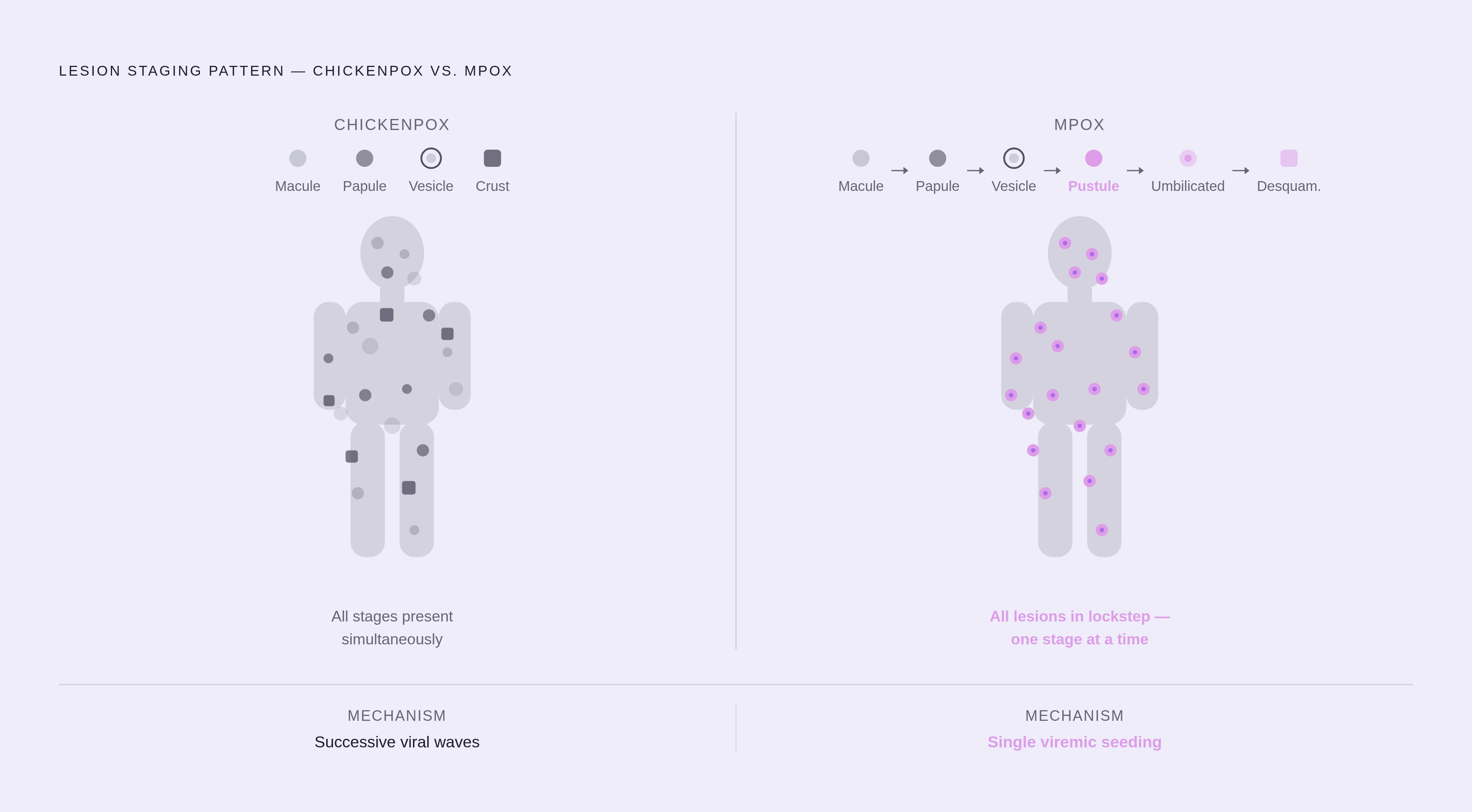
The visual signature, once you understand it, is actually quite specific: at any given moment, the lesions on the body are at multiple different stages simultaneously. One spot is still a flat macule. Another nearby has become a raised papule. A third is already a vesicle, and a fourth has crusted over. This mixed-stage picture, all phases present at once on the same patient, is the defining feature. The classic description of the vesicles is a "dewdrop on a rose petal": a small, clear blister sitting on a red base. That image is useful and not just poetic.
Distribution follows a pattern. The rash typically begins on the trunk and face, then spreads outward toward the limbs, and it spares the palms and soles. That detail becomes a differentiating point when comparing chickenpox to hand, foot, and mouth disease, which specifically involves those areas.
Before the rash appears, most patients experience one to two days of low-grade fever and malaise. Once the rash arrives, the itching tends to be relentless. Parents of young children who have been through this will confirm that description is not an exaggeration.
For otherwise healthy children, chickenpox is uncomfortable but self-limiting. For pregnant individuals and immunocompromised patients, the risk profile is substantially more serious. Lesions near the eyes, signs of bacterial superinfection such as spreading redness or warmth, and persisting high fever are all reasons to contact a clinician promptly rather than waiting it out.
Shingles: When the Virus Reactivates Along a Nerve Line
After chickenpox resolves, varicella-zoster virus doesn't leave. It retreats into the sensory nerve ganglia, where it can remain dormant for decades. In some people, usually older adults or those with diminished immune function, it reactivates. The result is shingles, herpes zoster.
The pattern is unlike anything else. The rash follows a single dermatome, the strip of skin innervated by one spinal or cranial nerve, and it is nearly unilateral. It does not cross the midline of the body. A blistering rash that wraps around one side of the torso like a belt, or runs down one side of the face, that dermatomal distribution is the single most recognizable feature. Nothing else looks quite like it.
Sequencing matters. Many patients experience several days of burning, shooting pain or heightened skin sensitivity in the affected area before any visible rash appears. This prodromal pain is often severe and can be confusing when the patient hasn't yet connected it to an impending eruption. There is also a variant called zoster sine herpete, where patients experience that same dermatomal pain without any visible rash ever appearing. I suspect more cases of unusual, localized neuropathic pain go unrecognized as zoster simply because there's nothing visible to prompt the right line of inquiry.
The treatment window is narrow. Antiviral medications, valacyclovir, famciclovir, and acyclovir among them, need to be started within 72 hours of rash onset to meaningfully reduce the severity and duration of the episode. This is not a situation for watchful waiting. Patients aged 50 and older, those with moderate-to-severe pain, and those with facial or ocular involvement are priority candidates for prompt treatment.
Postherpetic neuralgia, nerve pain persisting for months or years after the rash has healed, is a real and debilitating complication, disproportionately common in older adults. It's one of the primary reasons clinicians push vaccination hard for this age group.
Any involvement near the eye, herpes zoster ophthalmicus, is a same-day ophthalmology referral. Corneal damage from this variant can be serious and rapid.
Herpes Simplex: Cold Sores, Genital Herpes, and the Trigger Cycle
Herpes simplex virus exists in two primary types. HSV-1 predominantly causes oral herpes, the familiar cold sore. HSV-2 predominantly causes genital herpes. Both can infect either site, so the distinction is about prevalence, not exclusivity.
Cold sores are recurrent clusters of small vesicles appearing at or near the vermilion border, the edge of the lip where skin meets the red of the lip itself. Most people who get them learn to recognize the prodrome: a tingling or burning sensation that precedes visible blisters by 24 to 48 hours. That prodrome is the window when antiviral treatment is most effective, which is worth knowing practically. Consider what you felt before anything appeared, not just what you see — that's part of what you report.
Reactivation follows triggers, and patients tend to identify their personal constellation over time. Stress, febrile illnesses, intense UV exposure, and immune fluctuations are among the most commonly reported. Understanding your triggers lets you describe your outbreak history with more precision.
HSV enters skin directly through defects in the epidermal barrier. For patients with atopic dermatitis, where chronic inflammatory activity driven by IL-4 and IL-13 disrupts that barrier structurally, susceptibility to widespread HSV spread is meaningfully elevated. This can produce eczema herpeticum.
Eczema herpeticum is rare, but it belongs in a category of genuine dermatologic emergencies. When HSV spreads widely across eczema-affected skin, it presents as a sudden eruption of monomorphic, punched-out vesicles and erosions across areas already affected by eczema, sometimes with fever and signs of systemic illness. Here's what makes it easy to miss: the lesions emerge on skin that is already inflamed and disrupted, so the visual baseline is already abnormal. If an eczema patient develops a sudden, widespread, punched-out vesicular eruption, particularly with fever, that warrants urgent evaluation rather than reassurance.
For recurrent herpes simplex, oral antivirals can shorten individual outbreaks and, taken on a daily suppressive basis, reduce recurrence frequency. Worth discussing with a clinician if outbreaks are frequent or significantly disruptive.
Measles, Roseola, Rubella, and Fifth Disease: Four Rashes That Follow the Fever
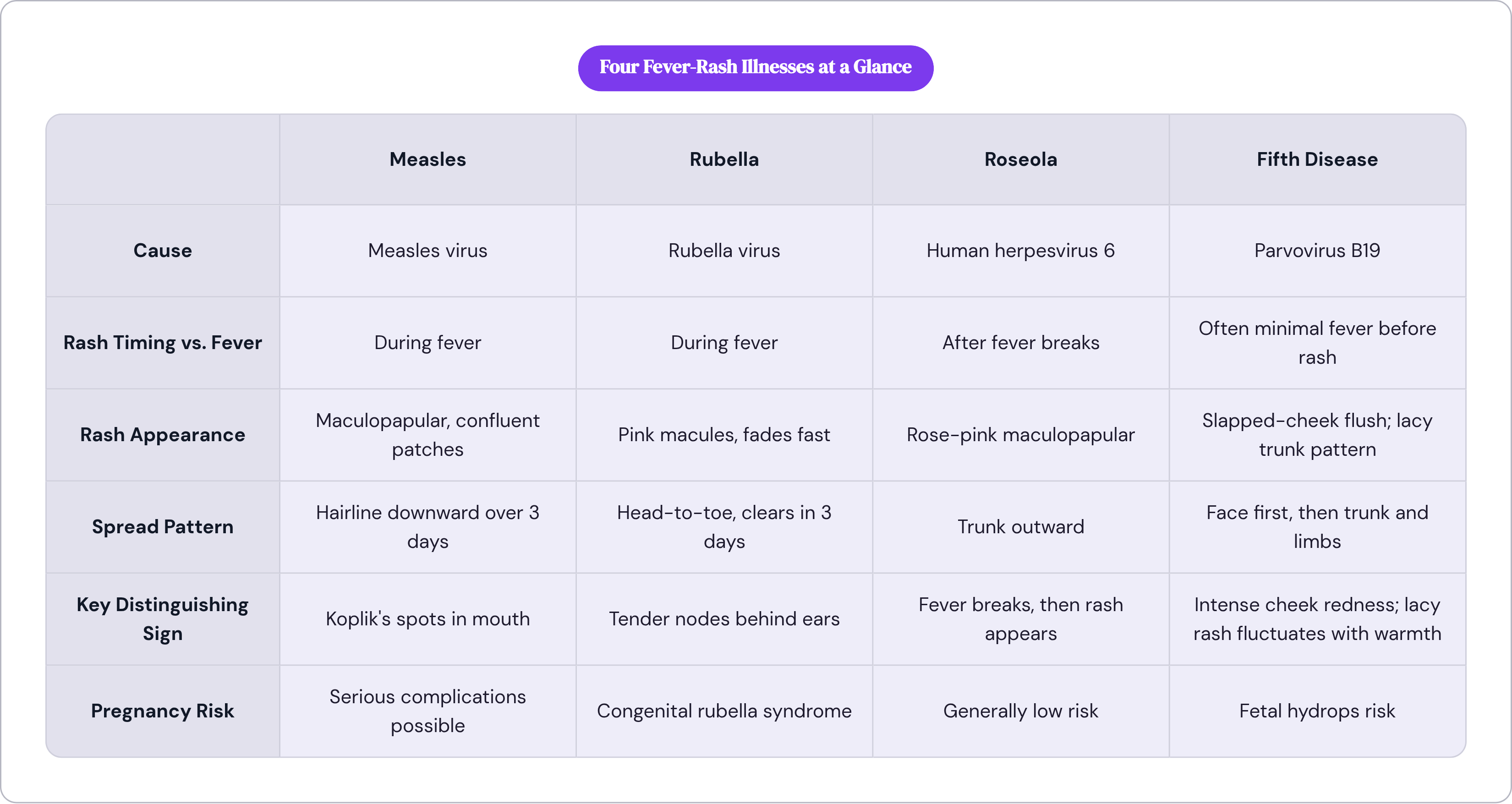
These four viral infections share a broad structural similarity: the rash arrives in temporal relation to a fever, either during it or as it resolves. Each, though, has a distinctive signature in timing, appearance, and distribution.
Measles
Measles announces itself before the rash appears. The prodrome is vigorous: several days of high fever, cough, runny nose, and conjunctivitis, sometimes called the "3 Cs." Not subtle.
Before the skin rash, look inside the mouth. Koplik's spots are tiny white lesions, often described as grains of salt on a red base, appearing on the buccal mucosa. They emerge a day or two before the skin rash and disappear as that rash develops. They are nearly pathognomonic, meaning their presence is a highly specific indicator of measles. This is the enanthem, and it's clinically valuable precisely because it arrives before anything visible on the skin.
The rash itself is maculopapular: flat and raised lesions together, starting at the hairline and behind the ears, then spreading downward across the face, trunk, and extremities over about three days. Lesions can coalesce into larger confluent patches as the rash progresses.
Measles has resurfaced in unvaccinated adult populations in recent years. Clinical suspicion matters even in adults who long assumed childhood illness vulnerabilities no longer applied to them.
Rubella
Rubella's rash is milder and moves faster than measles, though it follows a similar head-to-toe trajectory. The pink macules spread downward and often fade within three days. The distinguishing feature is lymphadenopathy, specifically behind the ears and at the base of the skull, notably tender.
In a healthy adult, rubella is often unremarkable. In a pregnant patient, it carries the risk of congenital rubella syndrome, with potentially serious consequences for the fetus. That changes the entire calculus of how aggressively it should be identified and documented.
Roseola
Roseola, driven by human herpesvirus 6, is most common in infants and toddlers. Its pattern is close to the inverse of the others: the fever comes first, often dramatically high, lasting three to five days, with no rash at all. Then the fever breaks abruptly. Then the rash appears.
That sequencing is the hallmark. The rash itself is a rose-pink maculopapular eruption beginning on the trunk and spreading outward, non-pruritic, typically fading within a few days. For parents watching a toddler run a high fever with nothing visible on the skin, knowing that a rash emerging precisely as the fever breaks is characteristic of roseola, rather than a sign of something worsening, is reassuring.
Fifth Disease
Fifth disease, erythema infectiosum, is caused by parvovirus B19. Its visual signature is probably the most distinctive of the four. The "slapped cheek" description is apt: an intensely red flush across both cheeks, demarcated and vivid, with relative pallor around the mouth. That facial rash is followed by a lacy, reticular pattern on the trunk and limbs that fluctuates with temperature, appearing more prominent when the child is warm and fading when cool.
Like rubella, fifth disease in a healthy child is typically mild. In pregnancy, parvovirus B19 can cause fetal hydrops, a serious condition. It doesn't belong in the category of "harmless childhood rash," and that distinction is worth making clearly.
The questions that begin separating these four: does the rash appear during the fever or after it? Does it start on the face and spread downward, or emerge on the trunk? Are the lesions discrete or do they merge? Are the lymph nodes involved? These aren't comprehensive diagnostics; they're the observations that narrow the field.
Hand, Foot, and Mouth Disease: Where the Lesions Appear Tells You What It Is
The name is its own diagnostic roadmap. Coxsackievirus A16 and Enterovirus 71 are the most common culprits, and the disease spreads with remarkable efficiency through daycares and schools.
The defining feature is distribution. Vesicular lesions appear specifically on the palms, the soles, and inside the mouth. That combination is the diagnostic signature. Palms-and-soles involvement immediately distinguishes it from chickenpox, which notably spares those areas. The oral ulcers are painful and often cause significant drooling and refusal to eat or drink in young children.
The skin lesions themselves are small, gray-white vesicles on a red base. They tend to be painful on the palms or in the mouth but not particularly itchy on the skin. Papulovesicular lesions can also appear on the buttocks.
The prodrome is brief: a day or two of fever, reduced appetite, and sore throat before the lesions emerge. The illness is self-limiting within seven to ten days, and treatment is supportive.
The concern to watch for is dehydration. When oral ulcers make swallowing painful, children can refuse fluids entirely, and dehydration develops quickly in small bodies. That's the most common reason hand, foot, and mouth disease escalates from manageable to urgent. Certain strains, particularly Enterovirus 71, have also been associated with rare but serious neurological complications, which is reason enough to take the diagnosis seriously rather than filing it automatically under minor pediatric annoyances.
Mpox: Uniform Lesion Progression and the Lymph Node That Sets It Apart
Mpox shares enough surface features with chickenpox that the two have been meaningfully confused, particularly as mpox became more widely recognized in recent years. The differences are specific and learnable.
The prodromal phase includes fever, headache, chills, sweats, sore throat, and muscle aches. That constellation overlaps with many viral illnesses. What separates it prodromally is lymphadenopathy, swollen lymph nodes. Lymphadenopathy is the pathognomonic sign separating mpox from varicella. Swollen lymph nodes accompanying a vesicular illness put mpox near the top of the differential.
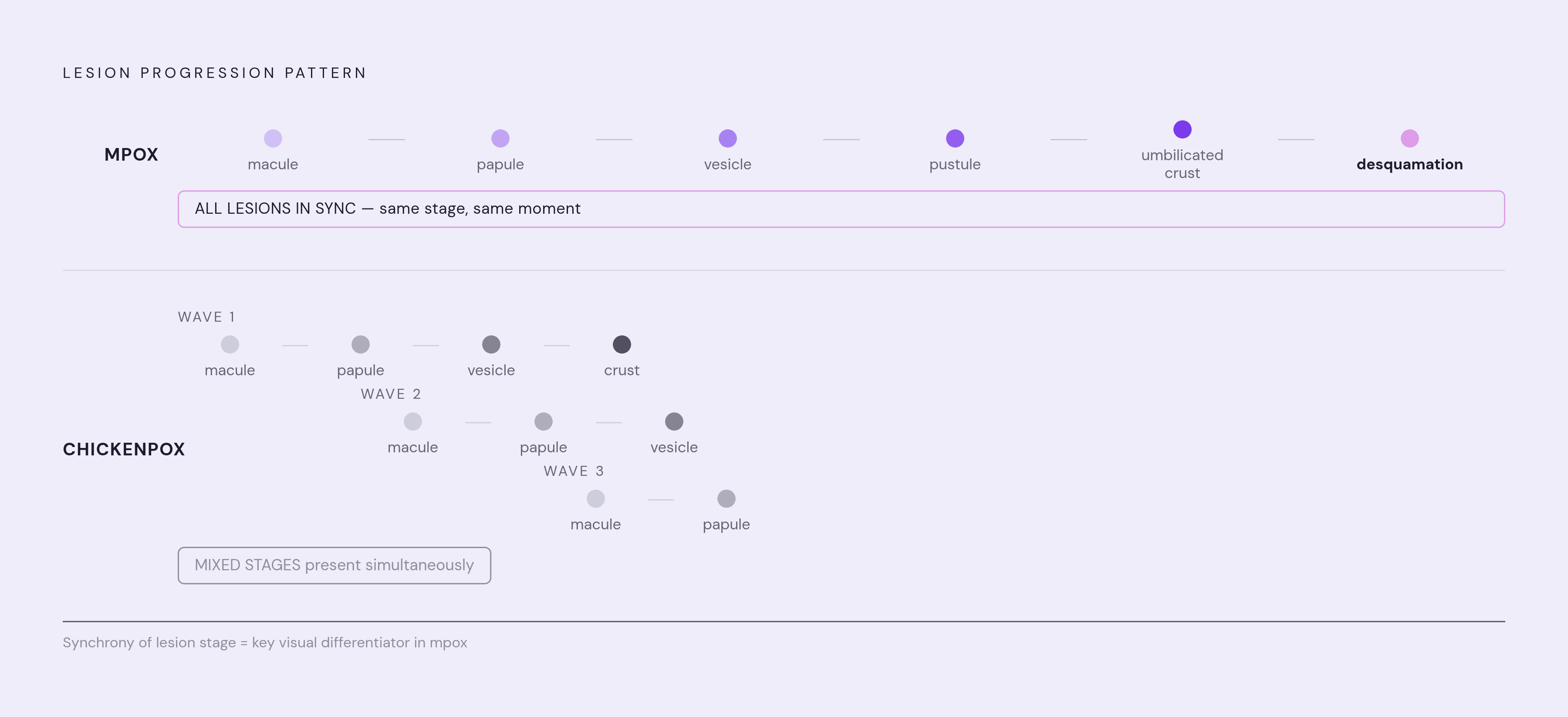
Lesion progression is the other defining characteristic, and the biology explains the pattern directly. In chickenpox, lesions are at mixed stages simultaneously: macules, papules, vesicles, and crusts all present at once across the body, because the virus arrives in successive waves. In mpox, all lesions evolve synchronously through the same stages at the same time, moving together from macule to papule to vesicle to pustule, then to umbilicated crust and desquamation. Mpox reaches the skin primarily through hematogenous spread during a single systemic viremic event, seeding lesions across the body at roughly the same moment. The biology produces the pattern; uniform progression is not an arbitrary distinguishing feature, it's a mechanical consequence.
The individual lesions in mpox tend to be deep-seated and well-circumscribed. Umbilication, the central dimpling, when visible, is a useful and specific visual cue.
Distribution has proven variable in recent outbreaks. The genital and perianal distribution seen commonly in recent years doesn't match the textbook images from older case series, which can lead patients to dismiss a presentation that doesn't look "classic." Location doesn't need to match a historical template to warrant evaluation.
What runs through all of these conditions, from childhood chickenpox to a shingles eruption in a 65-year-old to a toddler with hand, foot, and mouth disease, is that the skin is narrating something specific. The location, the lesion type, the timing relative to fever, whether lymph nodes are swollen, whether the lesions are all at the same stage or scattered across stages: each of those details is signal. You don't need clinical training to observe them. You need to know what you're looking for.