Deploying Dermatology-Specific Multimodal AI in a Clinical Setting Without Disrupting Existing Workflows
Dermatology AI only works in clinics when it fits existing workflows.
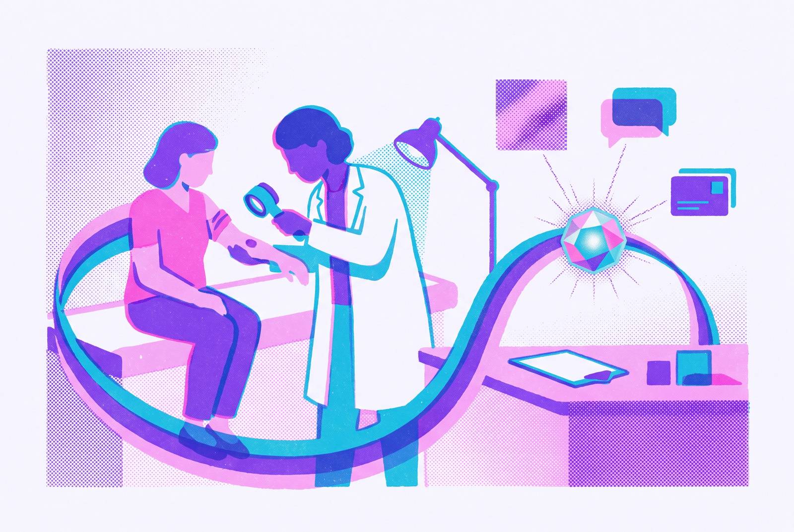
Dermatology-specific multimodal AI succeeds when its data inputs, outputs, and clinician review steps fit existing care workflows. PanDerm supports representation learning across several dermatology image types, while MEL-IA combines dermoscopic images with structured clinical metadata in a hospital-integrated system. These distinct capabilities leave the claim of a single “best” model trained on millions of labeled patient cases unsupported. Before deployment, an institution must weigh the training evidence and clinical scope against the data it can reliably supply and the results it can act on.
Why dermatology AI stalls after the pilot: the integration gap that model performance alone cannot close
Strong benchmark results do not automatically translate into bedside usefulness. Dermatology relies on what clinicians can see, image analysis is a large part of diagnosis, and large labeled datasets are available. Yet those advantages do not tell a clinic how an image will reach a model or where its prediction will appear. A pilot can show how well a model performs at classification while leaving both questions open.
Cheng et al. (2026) describe the progression from isolated image classification toward dermatology-specific multimodal clinical reasoning. That ambition requires more than a better classifier. Many clinics have no standard workflow for imaging, so the quality of images varies, as does the way metadata is captured. Those weaknesses can degrade performance before any algorithmic failure occurs. If routine care cannot supply the inputs a model expects, adding the tool around the workflow leaves the source of the problem intact.
What, then, separates a promising demonstration from routine use. The deployment has to connect acquisition, analysis, and clinical judgment without treating each as a separate project. Accuracy still matters, but it cannot fill in missing context or put a result in the clinical record.
Which multimodal AI tools support dermatology image diagnosis, and why is their data architecture more complex
“Multimodal” in dermatology means more than adding text to an image. Relevant inputs include clinical and dermoscopic images, total-body photography, dermatopathology images, structured medical histories, lab reports, multi-omics data, and longitudinal wearable signals. Each brings its own format, storage location, and access controls. The breadth creates an engineering burden as well as an opportunity. A model can combine evidence only if the system around it can reliably bring that evidence together.
PanDerm is a general-purpose multimodal dermatology foundation model that learns unified representations across total body photography, clinical, dermoscopic, and dermatopathology images. Earlier systems had a narrower scope, with a focus on single tasks such as skin cancer classification from dermoscopic images. Nahm et al. in JMIR Dermatology (2026) argue that credible local deployment must connect language models with vision models, including CNNs, vision transformers, vision-language models, dermatology-specific foundation models such as PanDerm, and medical multimodal models such as MedGemma. These names describe different roles.
For a buyer, that distinction limits what a shortlist can establish. The brief says which images PanDerm covers and describes MedGemma’s medical multimodal role, but gives no comparable labeled-case counts or clinical ranking. Meanwhile, dermoscopy images may sit in PACS, clinical metadata in HIS/RIS, and lab data in separate repositories. Each added input expands the integration surface. The choice of model depends in part on which of these sources the institution can bring together.
Interoperability as the foundational engineering requirement, not a secondary concern
Bringing those scattered inputs together as a clinical case that a clinician can use requires interoperability. Returning an AI output to the record where a clinician works also requires interoperability. Both directions are essential to the workflow. A system that retrieves images but leaves its result outside the clinical record has solved only part of the problem.
MEL-IA, described in J Med Syst (2026), shows how this works in a real hospital. It combines dermoscopic images and structured clinical metadata through a multimodal model. A mobile application supports structured image acquisition and metadata management at capture, addressing upstream variability. The deployment uses HL7 and FHIR for clinical messaging, DICOM and PACS for imaging, and HIS/RIS for hospital and radiology information. It achieved near-real-time processing and over 99% successful study integration.
The system’s evaluation also separates integration success from diagnostic evidence. External testing on HAM10000 found that its multimodal model outperformed the image-only baseline across most lesion classes, supporting the value of structured metadata. The paper calls for further clinical validation, and a working hospital deployment does not establish that implementation is cheap or easy for smaller practices. Nahm et al. describe complementary roles for FHIR in real-time exchange and the Common Data Model (OMOP) in retrospective interoperability across institutions.
Store-and-forward teledermatology as the deployment environment where AI integration pays off fastest
Store-and-forward teledermatology gives these connections a natural clinical sequence. Patients submit structured information and images asynchronously, so the case can be assembled before specialist review. That removes the need to synchronize patient attendance with the dermatologist’s assessment. It also creates a defined place for AI pre-analysis. Rather than interrupting a consultation, analysis can occur between submission and review.
Schramm, Haller, and Greis at the AI in Dermatology Summit 2026 describe an architecture that follows this sequence. Patient submission of history and images leads to AI pre-analysis for lesion detection, risk stratification, and triage. Dermatologist review follows, with validated escalation, before precision tele-monitoring through the EHR and digital therapeutics (DTx). The final stage extends the same architecture to chronic conditions such as psoriasis and atopic dermatitis.
Geisinger provides a useful boundary on the claim. Its working asynchronous model illustrates a workflow foundation, with AI triage still to be added. Within the proposed AI-assisted layer, large language models can support clinical summarization, structured data extraction, and differential diagnosis support, optionally grounded through retrieval-augmented generation. These functions reduce the unstructured burden of review. The benefit depends on presenting a coherent case to the dermatologist.
Local versus cloud hosting: a decision with privacy, governance, and equity implications that cannot be deferred
The location of that processing is a governance decision from the outset. In dermatology, protected health information includes written records as well as clinical photographs, dermoscopic images, and total-body photography. Such images may reveal identifiable anatomical features or document stigmatized conditions. Privacy cannot be separated from the data architecture. Hosting choices govern where this information goes and who controls changes to the model that handles it.
Nahm et al. make the case that local deployment has become more practical. Knowledge distillation, structured pruning, and low-bit quantization, combined with mixture-of-experts architectures, have lowered hardware requirements. Models in the 7- to 33-billion-parameter range can now run on consumer-grade workstations with modest NPUs or GPUs. This weakens the assumption that cloud dependency is unavoidable. It does not establish that every dermatology model or workflow will run adequately on such hardware.
Local hosting gives institutions control of clinical images and protection from vendor model deprecation that can disrupt validated workflows. It also permits auditing and fine-tuning on institutional schedules, including work on skin-of-color performance gaps. Control creates obligations as well as freedom. Heterogeneity across practices, model drift, and quantization-induced accuracy loss require structured mitigation through reporting frameworks including CONSORT-AI, SPIRIT-AI, DECIDE-AI, and TRIPOD+AI, together with approaches such as retrieval-augmented generation and federated learning. The paper does not argue for local-only deployment, because cloud models remain preferable for some tasks and the choice depends on institutional size, infrastructure, and workflow sensitivity.
Diagnostic equity as an integration requirement, not a post-deployment audit item
The ability to fine-tune locally becomes consequential when performance differs across patient populations. Dermatology AI systems perform worse on darker skin tones, and the problem starts with what is in the training data. Research on dermatology AI bias identifies dataset skew, with benchmarks including the ISIC dataset representing fair skin in approximately 70% of images. Darker skin tones are substantially underrepresented. A well-connected pipeline cannot by itself correct what a model has not adequately learned.
That distinction makes equity both a model requirement and a deployment responsibility. Assessment must check whether the people represented in training are like those who will receive care. Otherwise, a strong aggregate result can obscure a clinically important gap. The relevant outcomes extend beyond equal AUC to missed malignancies, delayed diagnosis, unnecessary biopsies, referral burden, treatment access, and patient trust. Assessment must look beyond summary metrics that look balanced to whether the system improves care for historically underserved groups.
A team deploying a system might reasonably ask whether researchers should do this work instead. Models do need better-balanced training data, but institutions that wait depend on what vendors choose to work on and when. The practical response described in the notes is to build local fine-tuning capability, federated learning across diverse populations, and institutional audit frameworks into deployment. These approaches give the institution ways to address gaps itself.
Human-in-the-loop as a workflow feature, not a safety disclaimer bolted on after the fact
A multicenter randomized clinical vignette study found that clinicians’ diagnostic accuracy meaningfully decreased when they saw systematically biased AI predictions. The finding exposes both behavioral and technical failure. When outputs are accepted uncritically, AI can worsen decisions. Oversight needs to ensure that clinicians check a result as well as see it.
Generative AI makes this challenge more acute because fluent, coherent language can sound better supported than it is. Users may give the confidence of the wording more weight in calibrating trust than the validation of the underlying model. Nahm et al. argue that locally hosted LLMs can require clinician acknowledgment before triggering downstream action. That turns oversight into a system constraint. Escalation criteria and transparent explanation outputs also need to be available where clinicians make decisions, as well as in a policy document.
Institutions should not wait for regulatory deadlines to build these controls. The EU AI Act’s high-risk obligations under Annex III were pushed back to December 2027, while Article 50 transparency duties apply from August 2026. The machine-readable marking obligation alone has a grace period until December 2026 for systems already on the market before August 2026. The AI Liability Directive was withdrawn from the Commission’s work programme in February 2025, with no direct fault-based successor announced, although an alternative remained possible. The revised Product Liability Directive (EU) 2024/2853 applies to AI software from December 2026, leaving institutions with governance responsibilities that cannot be reduced to a single compliance date.
What a workflow-first integration framework looks like in practice: four components that must cohere
The framework starts with a multimodal AI engine chosen for a specific clinical task. Representative validation data, accuracy, calibration, and escalation thresholds need to be established before deployment. Scope guides the model choice. PanDerm’s foundation-model approach and MEL-IA’s task-specific architecture serve different needs, and neither removes dependence on reliable capture. The modalities that are available must shape the choice of model before implementation begins.
The second part is standards-based clinical integration, which links those inputs to the clinical record. HL7, DICOM, PACS, FHIR, and OMOP support different parts of that connection, from live care to retrospective analysis. Integration has a clinical purpose. It must put the result at the decision point where it can be used, while supporting the data flows needed for continuous improvement. A technically successful transfer is insufficient if the output remains detached from care.
The third component is enforced human-in-the-loop validation through clinician acknowledgment gates, escalation criteria, and transparent explanations. The fourth is continuous learning and governance, including federated learning across diverse populations, reporting frameworks applied to updates, and audit trails for detecting drift before it affects outcomes. These components depend on one another. A model update changes what needs validation, while an audit is useful only if the institution can respond to its findings.
Asynchronous AI-assisted platforms that clinicians supervise offer a sound way to bring the four together. Patients submit structured data and images, AI pre-analyzes and risk-stratifies the case, and a licensed clinician reviews and decides. The architecture gives each responsibility a defined place. Intake, analysis, oversight, and follow-up can be designed as one workflow before a model is purchased.
Sources
- JMIR Dermatology - Local Deployment of Open-Weight Language Models in Dermatology: Viewpoint on Privacy, Equity, and Practical Implementation
- MEL-IA: An Interoperable AI System for Multimodal Skin Lesion Classification in Hospital Settings
- Meduniwien
- MEL-IA: An Interoperable AI System for Multimodal Skin Lesion Classification in Hospital Settings - PubMed
- Artificial intelligence (AI)-assisted diagnosis of skin diseases: From image classification to dermatology-specific multimodal clinical reasoning - PMC
- A Multimodal Vision Foundation Model for Clinical Dermatology