AI Diagnostic Tools That Fuse Dermoscopic Images With Patient History for Skin Disease Classification
Combining skin images with patient history helps AI distinguish look-alike conditions.

Multimodal clinical AI trained on labeled dermatology cases combines dermoscopic images with patient history to classify skin conditions using information that pixels alone cannot provide. Its advantage depends on paired image-and-history training data, useful clinical inputs, and clinician review. The strongest case for these tools is that they give clinicians better support when different conditions look alike.
Why dermoscopic image analysis alone is not enough for accurate skin disease classification
A melanoma and a dysplastic nevus can look alike under a dermoscope, yet distinguishing them has very different consequences for care. For an AI system, that creates a problem that sharper pattern recognition cannot always solve. If the visible evidence does not separate the conditions, the model needs information from outside the picture.
Dermoscopy still narrows the possible diagnoses compared with naked-eye inspection. But overlap persists. Certain variants of seborrheic keratosis, including adenoid or irritated types, can resemble pigmented basal cell carcinoma. Psoriasis and eczema can also share substantial dermoscopic features, particularly on the palms and soles, although their signatures are not fully identical. These examples show why visual similarity is not a reliable guide to whether two conditions belong in the same diagnostic category.
Could a larger image dataset resolve this problem? More examples can help a model learn visual distinctions that are present but difficult to detect. They cannot supply a lesion’s growth history, prior diagnoses, or medication history when those facts are missing from the input.
What clinical context actually contributes to a skin disease diagnosis
How likely is this condition in someone of this age? Spitz nevi are most common in children and young adults, though they can occur at any age. The same appearance can therefore raise different levels of concern for melanoma depending on the patient’s age.
The lesion timeline answers a different question. Is this a stable finding or a changing one? A lesion that grew over six weeks has a different prior probability from one that has been stable for a decade, even when their current images look identical. A snapshot compresses both histories into a single moment. Asking about change restores information that the image cannot retain.
Anatomical location helps establish which diagnoses fit the site. Acral lentiginous melanoma appears almost exclusively on the palms, soles, and nail beds, so location is part of the diagnostic evidence. Skin phototype and tone provide another frame for reading the image. Dermoscopic features read differently across Fitzpatrick skin types, and some pigmented structures need to be interpreted against the patient’s baseline pigmentation.
Symptoms and medical history add evidence about experience and risk. Does the lesion itch, bleed, or hurt? Bleeding without a history of trauma conveys something that a static image cannot explain, while medication and immunosuppression history can shift the probability of certain malignancies. Patients taking immunosuppressants have a sharply elevated risk profile for some cancers. A finding that appears low-probability in an immunocompetent patient may therefore warrant a different level of clinical concern in someone who is immunosuppressed.
How multimodal fusion architectures combine image features with structured clinical data
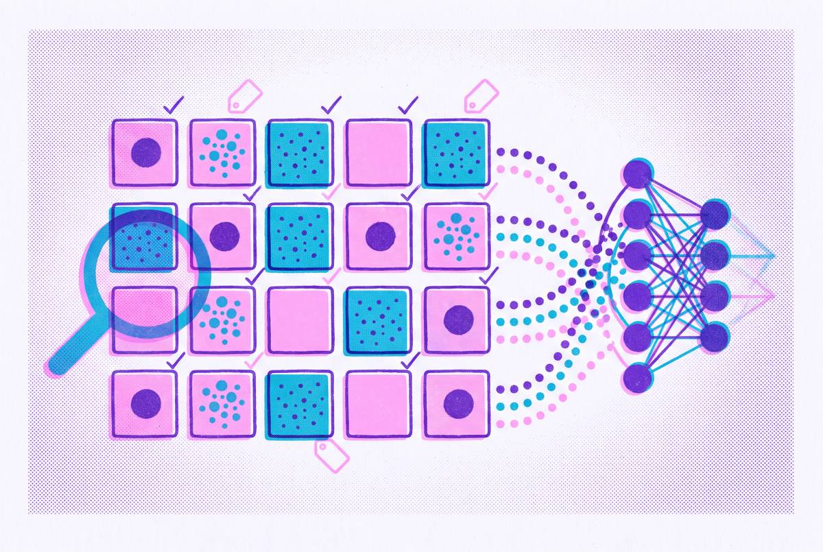
Fusion architectures bring these clinical variables into the classification process itself. The aim is to learn a joint representation in which visual features and clinical information both shape the result. This distinction is central to multimodal clinical AI trained on labeled dermatology cases. The model must receive both kinds of evidence and learn how they relate.
Early fusion combines image features and clinical variables before the main learning layers. The model sees the inputs together and must learn their relationships from scratch. Late fusion keeps the processing separate for longer. Separate encoders process the image and history, and their outputs meet at the classification stage through averaging, a weighted sum, or a learned gate. This approach is cleaner to train, but it limits how much the modalities can influence each other during feature learning.
Intermediate fusion allows that interaction to happen earlier. Cross-modal attention lets the tokens for image features and those for clinical text or structured data attend to one another as the model learns its representation. In plain terms, the model can learn that a visible feature has a different significance depending on age or lesion location. History helps interpret the visual evidence, rather than merely adjusting a finished image prediction. This makes intermediate fusion a more capable approach to learning context-dependent patterns.
Vision Transformers, or ViT, are well-suited to this design because their attention mechanism works with image patches. Clinical tokens can join the same sequence, so history and image patches share a representational space. Large multimodal models, or LMMs, extend the inputs further. They can take in free-text clinical notes, descriptions of symptoms, and patient-reported history along with images.
If the training objective rewards only overall accuracy, a model may rely on strong image signals and neglect history, failing on the ambiguous cases where context is most useful. Training must reward the use of history to resolve that ambiguity. It also requires paired cases containing images and history together, which are harder to collect than images alone. Because this training needs paired data, partnerships with clinicians matter, and training on images alone cannot take their place.
Where fusion models measurably outperform image-only models and where the gap closes
Fusion models have the biggest advantage when conditions look so much alike that image-only systems have more than one plausible classification. In these cases, informative history can produce a substantial improvement because it supplies evidence the visual model never received. Where the image already contains a clear distinguishing pattern, additional context may provide only a marginal lift. The useful comparison is therefore not just which model scores higher overall, but which kinds of cases account for the difference.
That distinction also limits what a broad accuracy claim can establish. A model’s success on visually clear cases does not demonstrate that it uses history well on difficult ones. Likewise, a small overall improvement can obscure a more consequential benefit within an ambiguous group.
A tool could handle low-ambiguity cases largely through image evidence while escalating high-ambiguity presentations to full fusion analysis. That approach can combine efficiency with accuracy and reflects how experienced dermatologists draw on additional context when appearance leaves uncertainty. It does not mean that clinical information is irrelevant to apparently straightforward cases. Rather, the amount of information needed to distinguish conditions varies with the presentation.
Rare conditions create a separate constraint. History helps only if it is informative and the model has learned enough from relevant paired cases to use it. A system that is well-calibrated for common conditions may remain uncertain about a rare genodermatosis despite receiving a detailed history. Claims about large labeled datasets require a clear account of paired clinical data and the conditions represented.
What fusion-based diagnosis requires from the patient side to work well
For patients, the practical implication is that describing the condition is part of supplying diagnostic evidence. A good image does not complete the input. When the lesion first appeared, whether it has changed, and how quickly it changed are among the most consequential details. Itch, bleeding, pain, and crusting add further context, as does a precise description of location. The history should also cover prior skin cancers, immunosuppression, sun exposure, and a family history of melanoma.
Why does precision make such a difference? A vague description leaves the model with an underspecified history, even when a history field has technically been completed. Saying that a lesion has changed without describing the timing gives less useful evidence than a clear account of its course. On ambiguous cases, missing details can push performance back toward that of an image-only model.
Clinician-designed structured intake forms help close that gap. They ask for variables the model needs rather than relying entirely on what a patient happens to include in an open-ended text box. Patients may describe what seems most noticeable while omitting something more diagnostically useful. Guided questions make those less obvious details easier to capture.
This supports asynchronous, message-based telehealth consultations built around guided intake. A well-designed intake can gather the context needed for AI-assisted triage without requiring a synchronous visit. The benefit depends on the questions. Patients are more likely to give accurate answers when they know why they are being asked about timing, symptoms, and medical history. Here, health literacy has a direct role in the quality of the evidence available for classification.
Why clinician oversight remains the essential layer above any fusion model's output
Even a well-trained fusion model produces probabilities. A licensed clinician must interpret those probabilities in the full context of the patient before deciding whether to biopsy, refer, or reassure. Fusion can improve sensitivity and specificity, but it does not eliminate false negatives. A missed melanoma has serious consequences, so errors do not all have the same clinical cost. A clinician still needs to review flagged cases even when a model does well overall.
Performance can also vary across patient groups. Darker skin tones, pediatric populations, and immunocompromised patients may be underrepresented in training data, leaving a model miscalibrated for those groups. Adding history does not automatically correct that imbalance. Clinician oversight provides a way to catch systematic errors that internal model metrics may not reveal. The full clinical context remains important precisely because the model’s learned representation may be incomplete.
Good decision support gives the clinician a better-structured problem. The model narrows the possible diagnoses, flags high-probability cases for urgent attention, and identifies the clinical variables it weighted most heavily. The clinician can then interpret that result. Differences between clinical judgment and model output provide a feedback signal for improving future model versions, making clinician involvement part of the system’s development as well as its use.
In asynchronous telehealth, that division of work has a concrete form. AI-assisted intake and preliminary assessment are reviewed by a licensed clinician before any care recommendation reaches the patient. The AI accelerates the review without replacing it. Together, these roles create a better system for reaching more patients with useful diagnostic support than either automated classification or unaided review alone.
Sources
- Exploring the Challenge and Value of Deep Learning in Automated Skin Disease Diagnosis
- JI-ADF: Joint-Individual Learning with Adaptive Decision Fusion for Multimodal Skin Lesion Classification
- Multimodal Machine Learning Approach for Diagnosing Atopic Dermatitis
- Validation of a Dermatology-Focused Multimodal Large Language Model in Classification of Pigmented Skin Lesions
- Artificial intelligence (AI)-assisted diagnosis of skin diseases: From image classification to dermatology-specific multimodal clinical reasoning - PMC
- Equity and Generalizability of Artificial Intelligence for Skin-Lesion Diagnosis Using Clinical, Dermoscopic, and Smartphone Images: A Systematic Review and Meta-Analysis
- Performance Evaluation of ChatGPT-4o in Dermatological Diagnoses Across Fitzpatrick Skin Types
- When AI and Experts Agree on Error: Intrinsic Ambiguity in Dermatoscopic Images