How Skin Changes Across a Lifetime
Newborns have structurally different skin that matures into adult form around age six.

Why newborn skin is not simply small adult skin
A newborn's skin is not a miniature version of adult skin. It is a structurally different organ at a different stage of development, and treating it like scaled-down adult tissue is where a lot of well-intentioned care goes sideways. The assumption that smaller means simply less developed is exactly the problem.
The barrier itself, the stratum corneum, the outermost layer of skin, forms during the last trimester of pregnancy and keeps adapting after birth. At delivery, it is thinner than it will eventually become, the individual corneocytes, the flattened dead cells that form the outermost skin layer, have a more irregular surface architecture, and the skin's pH sits higher than in adult skin. That insufficient acidification makes the surface more hospitable to pathogens and more vulnerable to irritants. It is one of the reasons diaper dermatitis and early atopic dermatitis show up when they do.
Within the first hours and days of life, skin gets colonized by commensal bacteria, the microbes that will form the skin's resident community. Which microbes arrive first depends heavily on how a baby is born. Vaginal delivery exposes skin to the mother's flora; cesarean delivery shifts that initial population toward skin and environmental organisms. That founding microbial community has downstream effects on how the immune system learns to calibrate itself, a process that plays out over years.
Sebaceous glands are actually highly active at birth, driven by maternal androgens still circulating in the newborn's system. Most people associate oil-producing glands with puberty, so this surprises them. Sebum levels peak in the first week and then fall sharply; the glands go quiet for years. But the machinery that reactivates at puberty is the same machinery, and that continuity matters more than it first appears.
What you're looking at in a newborn's skin is not abnormality. It is a biologically normal organ that is more permeable, more reactive, and more easily disrupted than it will be at any later point in life. Permeability is the thing to hold onto. It is both the source of specific vulnerabilities and the skin's opening position as it begins a decades-long process of structural refinement.
How the skin barrier matures through childhood and what can still go wrong
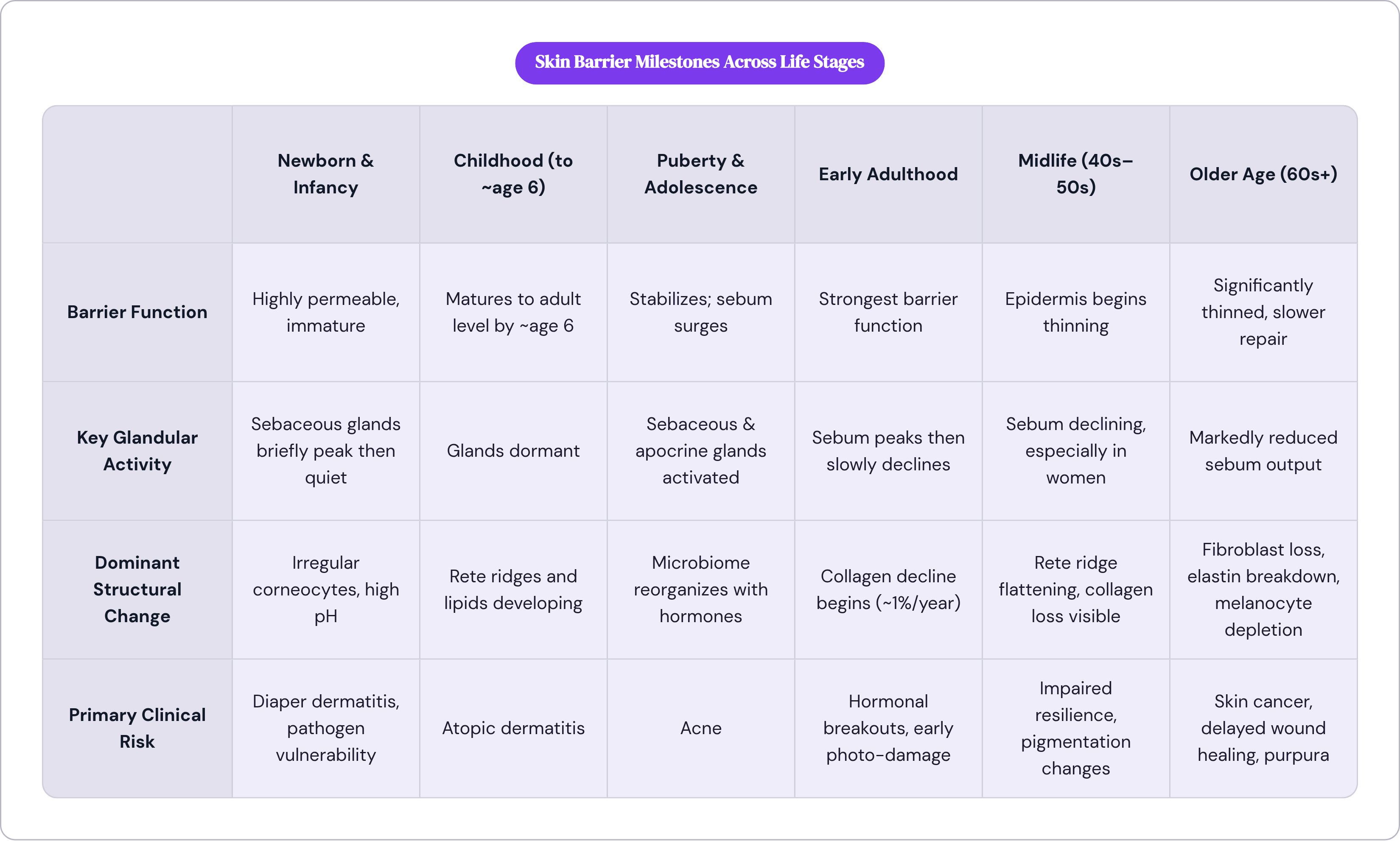
A 2023 study in Experimental Dermatology put a number to something clinicians had long observed qualitatively: the skin barrier reaches adult-level function, in terms of water loss, lipid organization, stratum corneum thickness, and corneocyte size, by around age six. Before that point, the barrier is measurably more permeable than it will ever be again in healthy skin.
Elasticity tells a related story. It rises from infancy to roughly age two, then plateaus, and the viscoelastic component, the skin's ability to return to shape after deformation, decreases progressively from infancy onward. The skin is most mechanically compliant very early and begins consolidating toward adult-range properties from there. Skin melanin content also increases across childhood, independent of sun exposure, so skin gradually darkens with age even before puberty introduces hormonal variables.
What disrupts childhood skin is mostly external: sun exposure, physical trauma, infectious illness. Measles, rubella, varicella; these are primarily skin diseases in terms of their visibility, but they are also where the skin announces something systemic. Intrinsic structural aging, the slow genetically programmed kind, is not a meaningful player at this stage.
The clinical story of childhood skin is largely the story of atopic dermatitis. The American Academy of Dermatology estimates it affects somewhere between ten and twenty percent of children in the United States, with most cases presenting before age five. An immature barrier, with thinner corneocyte layers and less compact lipids, is a more permeable barrier. A more permeable barrier lets allergens and irritants in more easily. The immune system responds. That cascade is the biology of atopic dermatitis in a sentence, and it happens precisely because the barrier has yet to reach the structural maturity it achieves around age six.
I've watched this get framed as failure, as a sign that something went wrong. That framing does real damage. The skin was temporarily operating below its eventual functional ceiling, and for some children, that window is long enough and porous enough to produce lasting inflammation. Reading the same biology that way changes what you do about it.
What puberty does to the skin and why the microbiome shifts matter
Puberty does not simply change what skin looks like. It changes what skin is.
Rising androgens trigger a surge in sebaceous gland activity. The glands that went quiet after the first week of life reactivate, and this time they stay on. Skin becomes oilier, particularly across the face and chest. Apocrine sweat glands, which were essentially dormant through childhood, are switched on by sex hormones. Their secretions are odorless until they make contact with skin flora, and then the odor begins. Body odor is a direct biological consequence of new glandular activity meeting a microbial community that is itself reorganizing, not a puberty side effect.
Research using Tanner staging, a standard scale for tracking puberty progression, documented something striking: the skin and nasal microbiomes shift dramatically between early childhood and adulthood, with the transition concentrated around puberty. The microbial community restructures in parallel with the hormonal one. These are not independent events. Treating them as separate phenomena leads to fragmented thinking about what adolescent skin actually needs.
Acne is where these threads converge in clinical practice. Excess sebum, a shifting microbiome, and alterations in the way follicles shed their lining cells; all three interact to produce the blockages and inflammatory responses that characterize acne. It is not a hygiene problem. Framing it otherwise, which remains common, does real harm to adolescents who are told that washing their face more carefully would solve it. The acne is a predictable biological outcome, not evidence of poor self-care.
What puberty actually establishes is a three-way relationship between sebum, the skin microbiome, and the barrier that will continue evolving for decades. In the teens and twenties it runs hot. In the thirties and forties it begins moderating. By the sixties it has largely reversed. Following that arc through adulthood explains why skin behaves so differently at each stage, and why interventions that work beautifully at twenty feel almost punishing at fifty.
The relative stability of adult skin and the slow losses already underway
The third and fourth decades are when you probably think of your skin as simply your skin. The barrier is as strong as it will get. Tone is relatively even. Healing is fast. There is no obvious reason to think structurally about what is happening underneath.
But the structural story is already moving. Collagen synthesis begins declining at roughly one percent per year after age twenty. In healthy young skin, the enzymes responsible for breaking collagen down are tightly regulated; collagen turns over slowly, with a full replacement cycle measured in decades. That regulation gradually loosens as the years accumulate, almost imperceptibly at first, then less so. Month to month, it is invisible. Compounded over twenty years, it is not.
Sebum production peaks in early adulthood and begins a slow decline in the fifth decade, particularly in women. Women's skin is also subject to cyclical hormonal variation across the menstrual cycle. The post-ovulation rise in progesterone relative to estrogen triggers breakouts in some women; pregnancy amplifies this pattern, with elevated hormones increasing oil production and driving the hyperpigmentation changes, particularly on the face, that many pregnant women recognize.
These are not pathological responses. They are the skin expressing hormone receptor activity distributed across keratinocytes, fibroblasts, melanocytes, and hair follicle cells. Estrogen receptors in these cell types actively regulate collagen synthesis, epidermal lipid production, and water retention. Knowing that explains why the hormonal transitions that come later produce skin changes as pronounced and as rapid as they do. The adult decades are the setup for everything that follows: the surface presents as stable while the structural scaffold is quietly contracting.
How the epidermis visibly thins and slows from the forties onward
At some point in the forties, the timeline that was running quietly in the background begins to surface. The epidermis, which had been losing roughly six percent of its thickness per decade, starts producing effects you can see and feel, sometimes quite abruptly.
One of the earliest and most consequential structural changes is the loss of rete ridges, the finger-like projections that interlock the epidermis (outer skin layer) with the dermis (the layer beneath it). As those ridges flatten, the dermal papillae that carry capillary loops delivering nutrients to the epidermis also attenuate. The epidermis is being progressively underfed by a structural interface that no longer holds as much contact surface, rather than failing outright. The mechanism matters because it changes what you can actually do about it.
Lipid production declines alongside this. Moisture retention drops, and when the barrier is disrupted, it takes longer to restore. Epidermal cell turnover slows, which means that damage, whether from irritation, a minor wound, or environmental insult, lingers longer than it used to. Skin in the forties is not fragile, but it is less resilient. The automatic recovery that characterized the thirties is still available; it just takes more time.
Reduced resilience and actual damage are not the same thing. The absence of quick recovery tends to read as something being wrong when it is actually just a slower clock, and that misreading shapes both the anxiety around it and the clinical response.
What happens inside the dermis as collagen and elastin break down
If the epidermis is the part of the story most people recognize, the dermis is where most of the age-dependent structural change actually occurs. These two timelines, epidermal thinning and dermal breakdown, are not sequential. They are concurrent, each compounding the other.
Fibroblast numbers decline measurably over time; research has found that total dermal fibroblasts in subjects aged eighty and older were reduced by an average of thirty-five percent compared to those in their twenties. Fewer fibroblasts means less collagen output, and the collagen that is produced changes in character. Aged skin shows less fiber alignment, more non-enzymatic crosslinks, and lower mechanical stiffness. The matrix metalloproteinase activity that breaks collagen down becomes less regulated, producing coarser, less organized fiber architecture. Advanced glycation end products, which accumulate as a byproduct of normal metabolism, stiffen collagen fibers further.
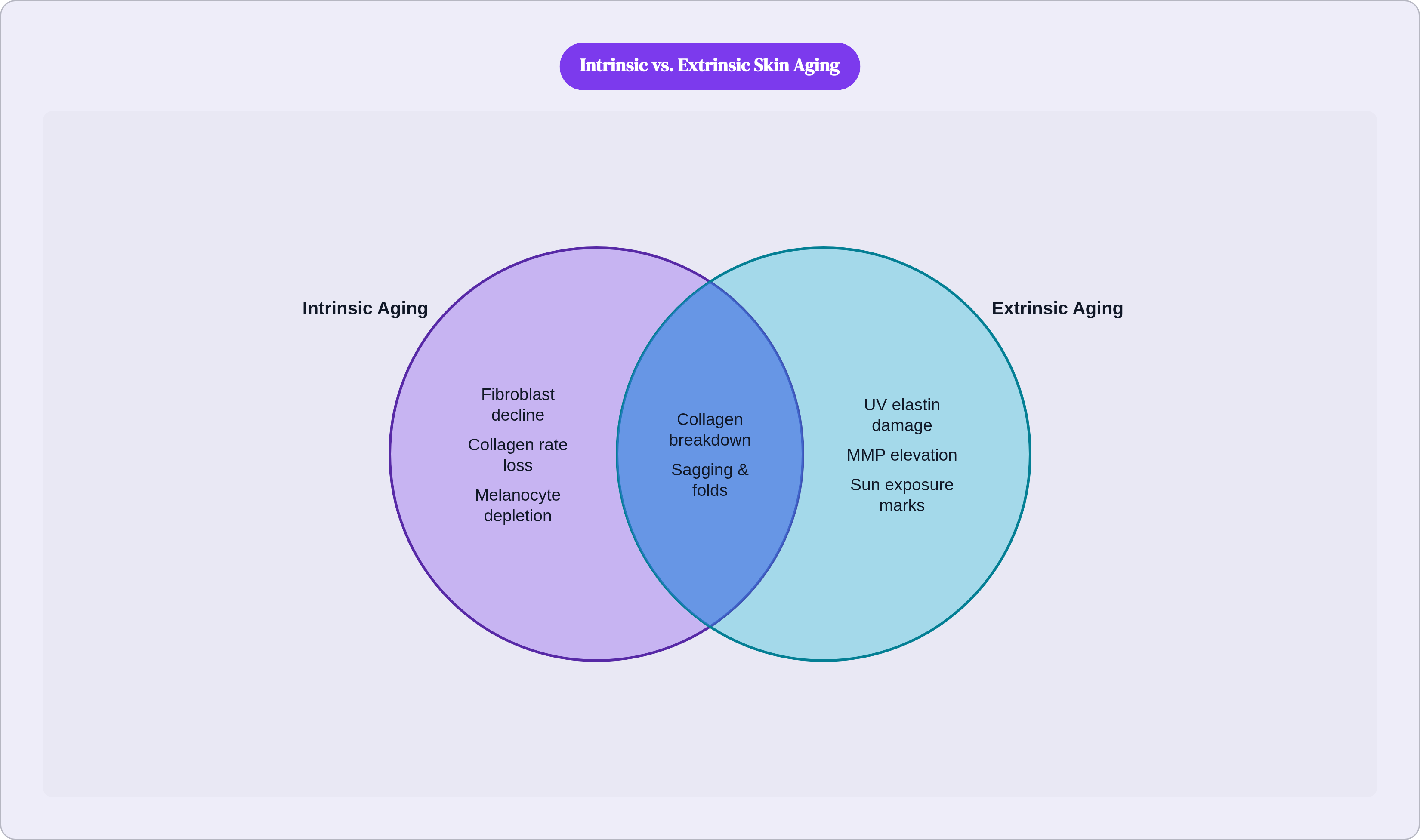
Elastin is degrading alongside all of this, and UV exposure accelerates its breakdown considerably. The combination of collagen and elastin loss is what produces the structural appearance most people associate with older skin: sagging, reduced rebound, deepened folds. Surface dryness contributes to the visual picture, but the architecture is the actual story.
This is also where the distinction between intrinsic and extrinsic aging becomes most legible. The slow, genetically programmed decline in fibroblast number and collagen output is intrinsic. The matrix metalloproteinase (MMP) elevation driven by UV, the accelerated elastin degradation from decades of sun exposure: those are extrinsic. The same skin is living both timelines simultaneously. By the time the effects are visible, they have been compounding for twenty or thirty years, which is part of why the changes, when they arrive, feel disproportionate to anything that happened recently.
How melanocyte loss disrupts pigmentation and immune surveillance simultaneously
The cells that produce melanin decline at a rate of roughly six to eight percent per decade after age thirty, with some estimates suggesting a ten to twenty percent reduction in functional melanocytes per decade. The numbers differ by source, but the direction is consistent and the implications extend well beyond the cosmetic.
Melanin is the skin's built-in UV filter. Fewer functional melanocytes means reduced photoprotective capacity, even in deeply pigmented individuals, even in people who have spent little unusual time in the sun. Because melanocytes deplete unevenly across the skin surface, and because cumulative UV exposure triggers erratic hyperactivity in the ones that remain, the result is not simply lighter skin. It is mottled skin: hyperpigmented spots alongside hypopigmented patches, an uneven distribution that reflects decades of uneven cellular stress. The geography of that unevenness tells a story about cumulative exposure, if you know how to read it.
Langerhans cells, specialized immune cells that patrol the skin, decline in parallel with melanocytes. Their function is immune surveillance: identifying foreign antigens, responding to pathogens, flagging abnormal cells. As their numbers and function diminish, the skin's ability to detect early malignant change is also diminished.
These two processes, pigment disruption and immune decline, share a stage in aging skin. If you are over sixty and notice a new spot, or one that is changing, it is worth having it examined rather than assuming it is nothing. The appearance and the biology are telling the same story, and the story is not purely aesthetic.
What menopause does to skin that most women are not told about
Estrogen does not affect skin as a side effect of other things it is doing. It actively regulates collagen synthesis, epidermal lipid production, and water retention through receptors distributed across the skin's major cell populations. When ovarian estrogen production drops sharply during menopause, the skin loses a regulatory signal that has been quietly maintaining its structure for decades.
The changes feel sudden in a way that is genuinely disorienting, precisely because they compress what had been a slow gradient into a much shorter window. Women often describe it as their skin changing faster than they expected, and that perception is biologically accurate. The rate of change accelerates in the perimenopausal and early postmenopausal period in ways that are now well documented.
The range of symptoms is wider than most women are told to anticipate. Dryness and itching get mentioned. Less commonly discussed: impaired barrier function, acne, rosacea, hair thinning, and increased facial hair. These are not separate conditions that happen to coincide with menopause. They are the skin expressing estrogen loss through the same receptor pathways it has been using all along. Framing them as unrelated events leaves women managing each symptom in isolation when the underlying driver is the same.
After ovarian estrogen production ceases, peripheral tissues, including the skin itself, begin compensating by converting other hormones into estrogen locally, using an enzyme called aromatase. The skin becomes an active endocrine site, not merely a passive target. That has real clinical implications for how skin-directed therapies work, and it represents a meaningfully different understanding of what menopausal skin is actually doing.
Clinical guidelines do not currently support hormone replacement therapy used solely for skin outcomes, because skin-specific trial data remains insufficient. That is a real limitation in the evidence. It also means that many women are managing significant, predictable, hormonally driven skin changes without a clear clinical framework for understanding them, uncertain whether what they are experiencing is normal aging or something worth raising with a physician. These changes are predictable and well documented in the research literature. They remain under-discussed in clinical settings, and that gap leaves women making decisions about their skin with considerably less context than they deserve.
How skin in older age becomes functionally vulnerable, and what is normal versus what signals a problem
By the mid-sixties, the processes running since the twenties have compounded enough to change how skin functions in daily life. The epidermis is thinner. The subcutaneous fat layer has thinned. The structural scaffolding of the dermis has lost significant volume. Sensitivity to touch, pressure, heat, and cold diminishes as nerve endings are affected. Blood vessels near the surface become more fragile, meaning that minor contact, a bumped arm, a sleeve sliding up, produces bruising or purpura without any force that would have registered as significant decades earlier.
Wound healing slows at every phase: the inflammatory response, proliferation, new vessel formation, re-epithelialization (regrowth of the skin surface), and tissue remodeling all take longer. Delayed healing is not defective healing. The outcome is slower; the mechanism is not broken. Treating slower recovery as pathology leads to interventions that are unnecessary and occasionally harmful.
Senescent cells add a layer of complexity. These are damaged cells that have stopped dividing but remain metabolically active, secreting inflammatory signals that interfere with surrounding tissue. They accumulate in aging skin. Research in animal models has found that clearing senescent cells accelerated wound healing and activated collagen-related genes. The translation to human skin is not yet established, but it points toward a mechanism researchers are beginning to treat as a serious therapeutic target.
The skin cancer statistics for this period are significant. Melanoma incidence and mortality rise sharply after age sixty. The majority of lifetime UV exposure happens in adulthood, not in childhood, which means the accumulation is largely invisible until it is not, and the consequences arrive decades after the exposures that caused them.
The full arc takes a while to see clearly. The collagen missing at seventy started declining at twenty. The melanocytes absent at sixty-five began depleting at thirty. What looks like sudden deterioration is almost always the long-delayed surface expression of processes that were underway quietly for half a lifetime. Recognizing that continuity does not eliminate the changes. But it shifts the question from what went wrong to what is worth watching, and in clinical practice, that is not a small shift.


