The Role of Melanin in Skin Tone and Protection
Eumelanin provides only SPF 13.4 at best, leaving all skin tones vulnerable to UV damage.

The two types of melanin that actually matter in human skin
Here is the fact that stops people mid-conversation: every human being, regardless of skin color, carries roughly the same number of melanocytes. The cells that produce melanin are distributed with surprising uniformity across all skin tones. What differs is how much melanin those cells produce, and what kind.
That distinction matters enormously, because melanin is not a single substance.
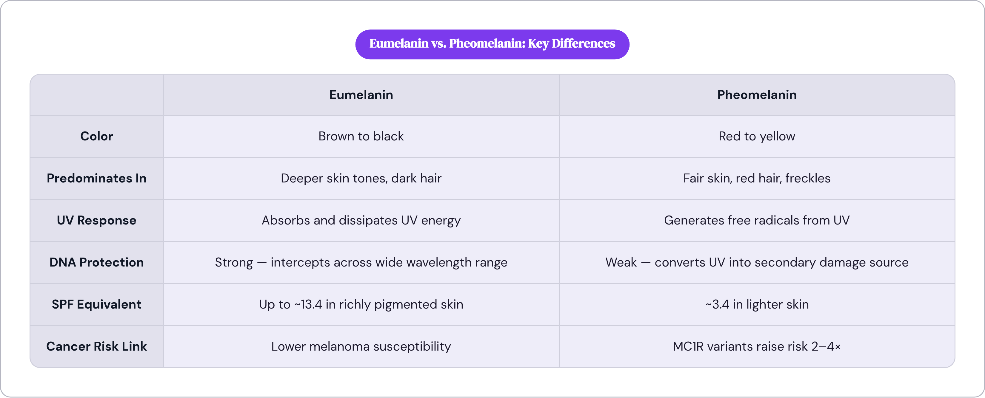
Five biological types exist. In human skin, only two are doing meaningful work. The first is eumelanin, the brown-to-black pigment predominating in deeper skin tones. The second is pheomelanin, the red-yellow pigment concentrated in fair skin, red hair, and freckles. Most people carry some blend of both, and that blend is where the real story lives.
Eumelanin does the heavy lifting. Its absorption spectrum, the range of light wavelengths it captures, increases exponentially as wavelengths approach the UV range, which is unusual behavior for a biological pigment. Most biological pigments work differently. Eumelanin intercepts UV energy across a wide range of wavelengths and dissipates it efficiently. Pheomelanin does something closer to the opposite: when UV strikes it, rather than absorbing and dispersing that energy, it generates free radicals. It converts the UV hit into a secondary source of cellular damage.
The ratio between these two pigments is primarily governed by variants in the MC1R gene, producing a continuous spectrum of UV sensitivity rather than any clean binary of light versus dark. How readily you burn, how efficiently you tan, what your baseline cancer risk looks like: all of it tracks that ratio. And everything downstream in this discussion depends on understanding it.
How melanin physically shields DNA from UV radiation
The word "pigment" undersells what melanin does. It suggests something passive, something that merely tints tissue. In practice, melanin functions as a dynamic defense system running at least three interlocking mechanisms simultaneously.
First, absorption: melanin intercepts UV radiation before it reaches the deeper cellular layers where the most critical biological machinery lives. Second, dispersion: it scatters UV energy to reduce DNA strand breaks. Third, neutralization: melanin acts as a free-radical scavenger, clearing reactive molecules before they can attack DNA.
The most remarkable feature of this system is what researchers call the supranuclear melanin cap, a concentration of melanin positioned directly above the nucleus of each skin cell. Melanin doesn't float around keratinocytes at random; it positions itself as a direct, geometric shield over the DNA. Researchers first characterized this arrangement in the mid-1990s. It is both denser and more prevalent in darker skin tones, and it suggests a level of organizational precision that the word "pigment" fails to honor.
Then there's the speed of the response. Research published in the Proceedings of the National Academy of Sciences documented that eumelanin converts UV energy into heat through a photochemical reaction occurring on a femtosecond timescale, a quadrillionth of a second, meaning the dissipation happens so rapidly that free radicals essentially lack the time to form. The cellular response cascades outward from there: limit UV penetration, produce protective metabolites during absorption, and if damage still slips through, activate DNA repair signaling. The system monitors and adapts rather than simply sitting there as a static barrier.
Which makes it all the more striking that the SPF equivalent numbers are as modest as they are.
What melanin's SPF equivalent actually means in practice
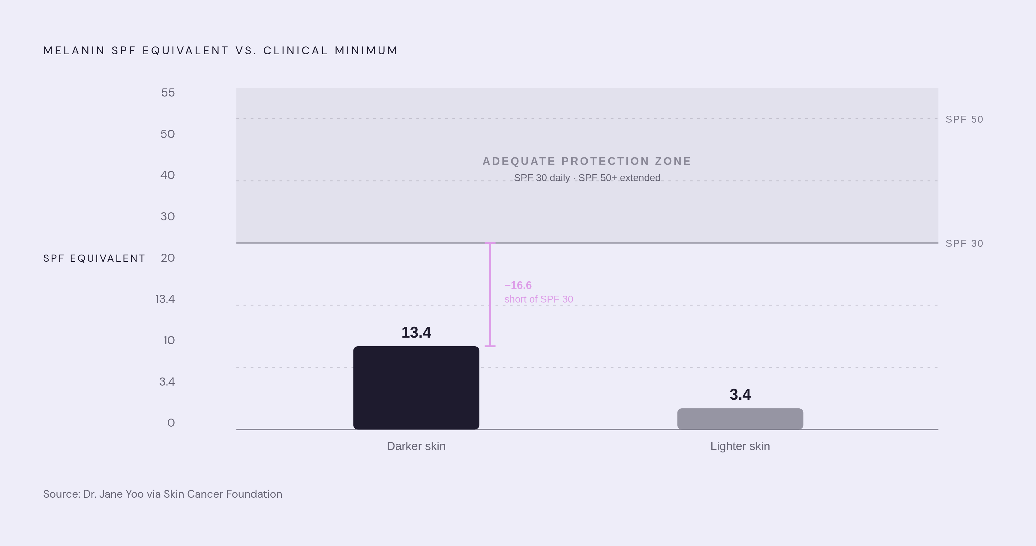
The figure that consistently surprises people: melanin in darker skin provides an SPF equivalent of approximately 13.4. In lighter skin, approximately 3.4. Those numbers come from dermatologist Dr. Jane Yoo, cited through the Skin Cancer Foundation, and they warrant a pause.
Neither figure clears the threshold dermatologists recommend for adequate daily protection. The clinical minimum for meaningful protection is SPF 30, with SPF 50 or higher for extended exposure. Eumelanin at its most robust doesn't get you there on its own.
There's also a wavelength dimension that matters practically. Eumelanin blunts UVB, the burning wavelength, more effectively than it blocks UVA. UVA penetrates more deeply into the dermis, drives accelerated photoaging, and generates indirect oxidative DNA damage. It's present year-round, passes through glass, and produces no immediate sensory feedback the way sunburn does. Damage accumulates quietly. This is precisely the wavelength sunscreen addresses best and that melanin handles least well.
Epidemiological data confirms the protection gradient as exactly that: a gradient, not a binary switch. Individuals at the fair end of the Fitzpatrick scale, a standard medical classification of skin tone from very fair to very dark, face meaningfully greater UV-induced damage and tumor formation risk. That gradient is real and significant. It is not the same as saying darker skin is fully protected.
Melanin modifies risk. It does not eliminate it. The distance between "less vulnerable" and "adequately protected" is exactly where sunscreen, regular monitoring, and skin literacy have to do their work.
The genetics that set a person's melanin baseline — and raise melanoma risk independently of sun exposure
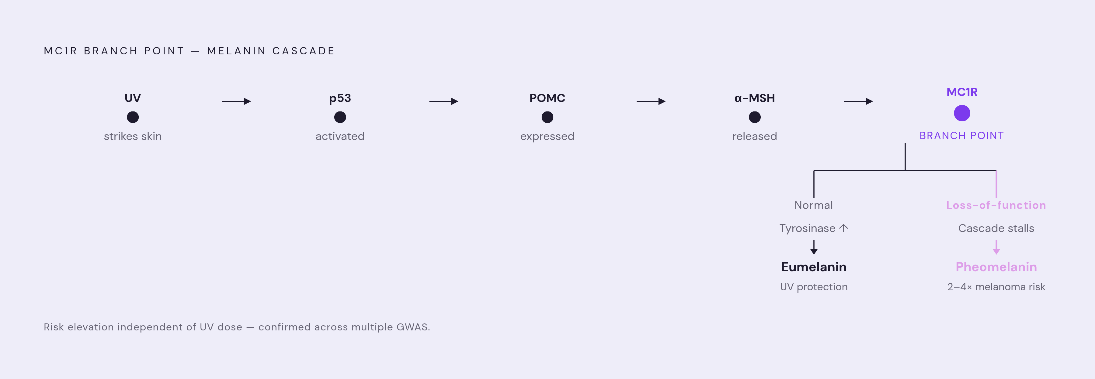
The MC1R gene sits on chromosome 16 and functions as the master regulator of the melanogenesis pathway, the biological process that produces melanin, including tanning response, eumelanin-to-pheomelanin ratio, skin and hair color, and melanoma susceptibility. When UV strikes the skin, it triggers a signaling cascade running roughly from p53 through POMC to alpha-MSH, which binds MC1R and upregulates tyrosinase expression, producing more melanin as a direct damage response. The pathway is elegant and several steps deep.
The clinically consequential finding: certain loss-of-function MC1R variants shift production toward pheomelanin and are associated with approximately a two- to fourfold increase in melanoma risk. Multiple genome-wide association studies across large populations have confirmed this, and the elevation is independent of UV dose. Two people with nearly identical sun histories can carry meaningfully different melanoma risk based on inherited MC1R variants alone.
That's worth sitting with. It means your sun behavior, while important, is not the only variable. It also means telling yourself "I've never really been a sun person" is an incomplete risk profile.
These variants run in families. If a parent or sibling has red hair, fair skin, and a history of melanoma, that combination is not merely cosmetic information. It belongs in a serious conversation with your doctor about sun protection, sunscreen selection, and how often you should be monitored, not treated as an afterthought.
The less-known side of melanin: when it can contribute to oxidative damage
Melanogenesis, the process of actually making melanin, is metabolically stressful. Melanocytes operate under constant oxidative stress because the synthesis process itself generates damaging molecules, superoxide and hydrogen peroxide, as byproducts. The cells responsible for your protective pigment are, in effect, under continuous low-level chemical assault from their own production machinery. I've found this to be one of the more unsettling elegances in human biology.
A more surprising pathway involves what researchers call chemiexcitation, a process where chemical reactions, rather than light directly, produce an excited molecular state that can damage DNA. When UV exposure activates nitric oxide synthase and NADPH oxidase, their products combine to form peroxynitrite, a reactive compound that oxidizes melanin into an unstable, high-energy state. That energized melanin then transfers energy directly to DNA, causing the same mutations UV normally produces, without any additional UV photons arriving. The damage, in this scenario, occurs downstream of the UV exposure, through the melanin that was supposed to be protective.
The carbonyl compounds formed in this process are reactive; they bond with DNA and proteins. Some researchers believe this pathway contributes to melanoma initiation, particularly in heavily pigmented cells under sustained oxidative stress.
This doesn't overturn the net protective picture epidemiological data shows across populations. But it does account for things melanin's standard protective narrative cannot explain: why melanoma develops in heavily pigmented individuals, and why the mechanism of damage is not simply a function of how much UV penetrates to begin with. Melanin, under the right conditions, participates in the damage it was supposed to prevent. That's a more complicated picture than most people carry around, and it's accurate.
Why darker skin has lower skin cancer rates but worse outcomes when cancer develops
Both sides of this epidemiological picture deserve to stand rather than letting one number absorb the other.
White individuals are approximately 20 times more likely to develop melanoma than Black individuals. That gap is real, meaningful, and largely attributable to melanin's protective effects.
And yet, five-year melanoma survival rates are significantly lower in Black patients. The American Academy of Dermatology has documented that 22% of melanoma cases in African American patients are diagnosed after cancer has already spread to nearby lymph nodes, and 14% after spread to distant organs. These numbers describe a systemic failure, not a biological one. The cancer is not more aggressive; it is found later.
Part of the explanation is anatomical. In darker skin, melanoma frequently appears on UV-unexposed sites: the palms, soles, groin, and oral mucosa. Standard skin cancer guidance orients people toward monitoring sun-exposed areas. Applied to darker skin, that guidance is incomplete at best, and the incompleteness has consequences.
A 2024 study examining roughly 1,200 online skin cancer images found that 95% depicted lighter skin tones while just over 1% depicted dark skin tones. That gap doesn't stay abstract. It shapes what patients recognize as concerning in their own skin, and it shapes what clinicians learn to detect during training.
Bob Marley died in 1981 from acral lentiginous melanoma, a type of skin cancer that develops on palms, soles, and under nails, which began as a dark spot beneath a toenail he had attributed to a soccer injury. His case gets cited repeatedly not because it is exceptional, but because it illustrates exactly this failure mode. A man who had no reason to think his skin required that kind of vigilance, because nothing in the cultural or clinical environment around him suggested it did.
The protective biology of darker skin creates a real advantage. It also generates a false sense of comprehensive protection, one that medical education and clinical imagery weighted toward lighter skin reinforce rather than correct.
The real trade-off between melanin and vitamin D production
The mechanism is fairly direct: sunlight converts a precursor molecule into vitamin D in the deeper dermis. Melanin, absorbing UVB in the upper layers, slows that conversion by competing for the same photons. More melanin means more UV protection and slower vitamin D synthesis. The same mechanism producing two opposing outcomes simultaneously, with no clean workaround available.
Current clinical standards identify ethnic minority populations, including Asian and Black individuals, as high-risk for vitamin D deficiency. That risk is documented.
Where the picture becomes less settled: recent research suggests melanin-rich skin utilizes distinct vitamin D metabolic pathways that produce active metabolites not captured by the standard 25-hydroxyvitamin D blood test. If that finding holds, measured deficiency in some darker-skinned individuals may overstate actual functional deficiency, because the test isn't measuring what the body is producing and using.
Whether current testing thresholds are appropriate for melanin-rich skin is contested. The practical implication is narrow but worth stating clearly: if you have darker skin, a vitamin D lab value is worth discussing with a clinician rather than acting on unilaterally. The number may not mean exactly what it appears to mean.
What melanin dysfunction looks like — and how pigmentation disorders signal underlying biology
Pigmentation disorders tend to get framed as cosmetic concerns. They are more usefully understood as diagnostic signals from the same pathway governing UV defense.
Vitiligo involves the autoimmune destruction of melanocytes, producing white patches across the skin's surface. It affects roughly one-half to two percent of the global population, typically presenting before age 30. When melanocytes are destroyed, so is the UV protection they provided, distributed unevenly across the surface they once occupied.
Melasma demonstrates a different dimension of the pathway: hormonally driven overproduction of melanin, frequently triggered by pregnancy or oral contraceptives and worsened by UV exposure. The melanogenesis cascade is sensitive to hormonal signaling, not only to UV, which is part of the system's design. It's also why certain life stages produce visible, unexpected changes in skin tone that seem to appear from nowhere.
Post-inflammatory hyperpigmentation, appearing after acne, eczema, or physical trauma, is the same repair signaling that guards against UV damage applied to a context where the original injury had nothing to do with UV. The biology is functioning correctly. The output is disruptive because the trigger was wrong.
Albinism, in its most complete form, involves the absence of functional tyrosinase, the enzyme that controls the pace of melanin production. Without it, the entire synthesis cascade stalls. Individuals with this condition have minimal melanin protection regardless of ancestry, with correspondingly elevated UV damage risk across every skin surface.
The same pathway runs through all of it: tyrosinase activity, melanocyte survival, melanosome transfer. Each disorder represents a malfunction at a specific node in a system that is otherwise working to protect you. Reframing pigmentation changes as information rather than cosmetic noise is how you start to read that system clearly.
How understanding melanin changes the way any skin tone should approach sun protection
The SPF 13.4 ceiling is the most practically useful number in this discussion. No skin tone reaches adequate protection from melanin alone, which means sunscreen isn't a product designed around fair skin that darker-skinned people should also consider using. It's designed around a protection gap that exists across every Fitzpatrick type, just at different depths.
If you have fair skin and known MC1R variants or a family history of melanoma, your elevated baseline risk is partly genetic, not purely behavioral. That shifts the calculus toward how often you get checked alongside your daily SPF selection. You can do everything right with sunscreen and still carry inherited risk that warrants more frequent clinical attention.
If you have darker skin, your self-examination needs to extend beyond sun-exposed surfaces. Nails, palms, soles, and mucous membranes: these are the sites where acral and mucosal melanomas present, and they are largely absent from standard skin check messaging.
How you interpret your vitamin D result should account for your skin tone and the genuine uncertainty about whether standard testing fully captures functional vitamin D status in melanin-rich skin. A number alone is a complete answer only when the test is calibrated to you.
Pigmentation changes, new dark patches, asymmetric spots, marks that persist without resolving, are signals from the same biological system governing UV defense. They warrant clinical attention.
Start with an accurate account of your own melanin picture. Your skin tone, family history, sun sensitivity, and pigmentation patterns are not background details. They are the foundation for every practical decision you make about protection, monitoring, and supplementation. Melanin is a remarkable system. It is not, by itself, enough.


