Common Benign Skin Growths and Their Characteristics
Most benign skin growths arise from normal aging of the epidermis, dermis, or subcutaneous fat.

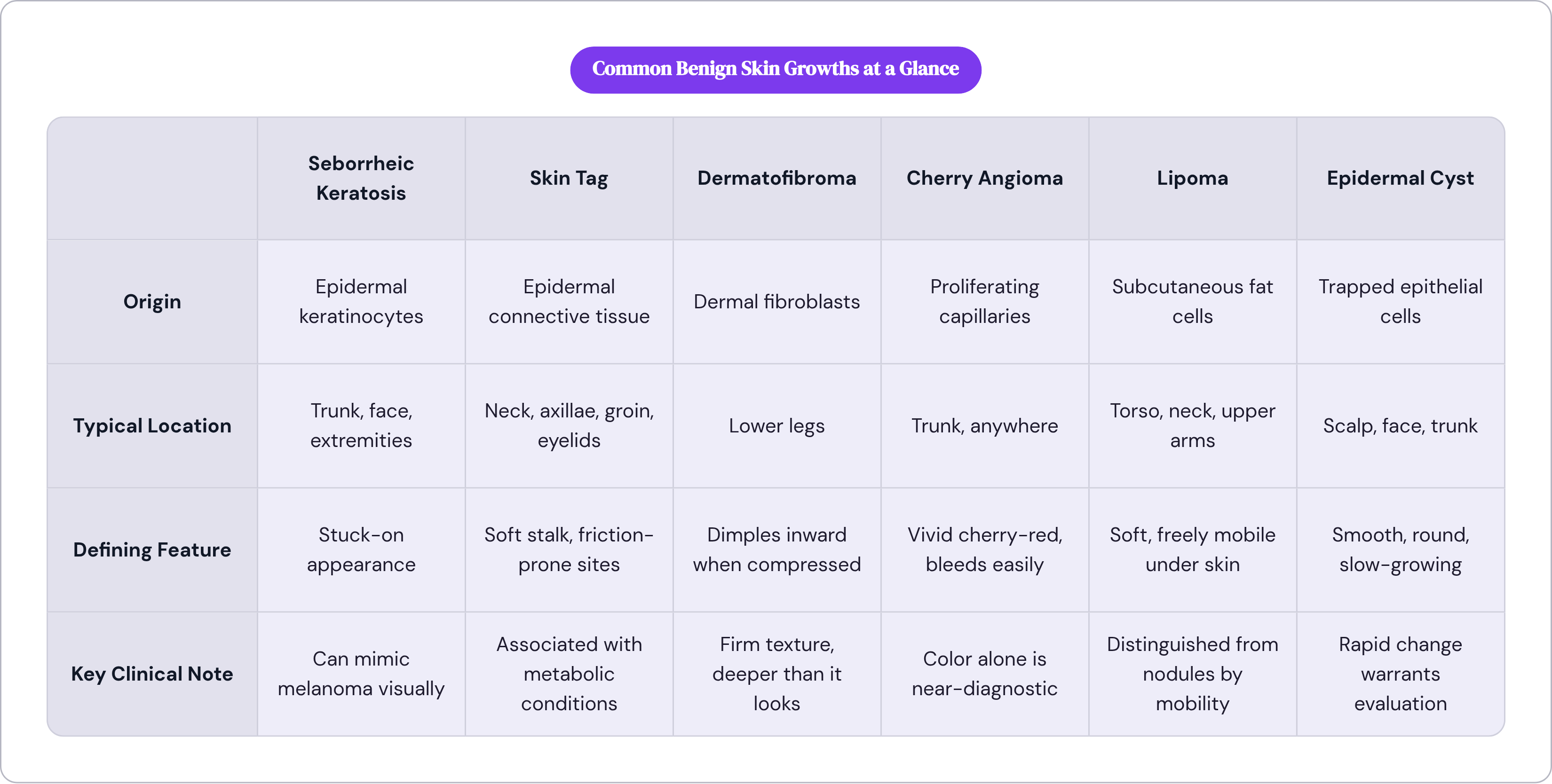
The skin has two primary structural layers: the epidermis on top, the dermis beneath it. The epidermis is built primarily from keratinocytes, organized into ascending sublayers from basal to cornified. The dermis houses fibroblasts, fat, and the capillary networks that supply the whole system. Benign growths correspond to those resident cell populations. Seborrheic keratoses arise from immature epidermal keratinocytes. Dermatofibromas come from fibroblast accumulation in the dermis. Lipomas develop from adipocytes in the subcutaneous layer. Cherry angiomas arise from proliferating capillaries. Cysts form when epithelial cells get trapped.
The basal layer, the deepest level of the epidermis, is where keratinocytes replicate continuously. To understand why this works, we must first look at what happens when daughter cells lose contact with the basement membrane beneath them: they receive a chemical signal to stop dividing and begin differentiating into mature cell types. When that signaling breaks down, even slightly, proliferation continues unchecked in a localized way. That mechanism underlies most benign epidermal growths.
Aging is the dominant driver. Research demonstrates a striking decrease in epidermal thickness when keratinocytes are co-cultured with an aging dermis, which helps explain why the prevalence of most benign growths tracks closely with advancing age. UV exposure and genetic predisposition contribute as well, but their relative weight varies by growth type. Sun exposure matters more for lesions with melanocytic or keratinocytic origins; genetic predisposition is a stronger factor for some cyst types and for multiple lipomatosis. Neither is irrelevant, and the proportions are complicated, which means anyone offering a single clean explanation is leaving something out.

Seborrheic keratoses: the most common benign skin growth most people can't name
If you asked a hundred people to name a common benign skin growth, almost none of them would say seborrheic keratosis. Yet this is, by a wide margin, the most prevalent benign epidermal growth in adults. It appears across a broad visual spectrum: a lightly pigmented, superficial patch on one end; a thick, dark, scaly plaque on the other. The hallmark that unites all of them is what clinicians call the "stuck-on" appearance, as though someone placed the lesion on top of the skin rather than growing it from within. Once you've seen that, you start noticing it everywhere. On strangers' necks in grocery store lines. On your own forearms, eventually.
Prevalence rises sharply with age. Among people in their mid-teens to mid-twenties, roughly twelve percent are affected. By the time a person is past sixty, the numbers approach universality; Australian data show an average of sixty-nine seborrheic keratoses in patients over seventy-five. This is not a rare finding. It is, in the most literal sense, a near-universal feature of aging skin.
A clinically important variant that receives disproportionately little attention in patient education is dermatosis papulosa nigra, which affects up to thirty-five percent of African Americans. It presents as smaller, darker papules predominantly on the face, and while it is histologically identical to typical seborrheic keratoses, it is frequently misidentified or overlooked in standard patient materials. Patients who are not told what to look for will miss the connection entirely. That gap in patient education is worth naming plainly.
Despite their benign nature, seborrheic keratoses can cause real functional problems. Lesions in friction-prone locations — belt lines, brassiere-strap areas — are prone to irritation, pruritus, and occasional bleeding. The issue is mechanical, not oncologic, but it is the most common reason patients seek treatment for growths they've otherwise tolerated for years.
The more pressing clinical challenge is visual variability. Seborrheic keratoses are variable enough to be confused with melanoma or other malignant lesions on naked-eye examination, and atypical presentations are not unusual. That mimicry problem will reappear throughout this piece, because it is central to understanding why even benign growths reward some level of visual fluency.
Skin tags, dermatofibromas, and cherry angiomas: three common growths with distinct signatures
Skin tags
Skin tags, clinically known as acrochordons, are soft, pedunculated papules connected to surrounding skin by a narrow stalk. They're typically the same color as adjacent skin or slightly darker, and their distribution follows a clear logic: they appear in intertriginous areas where skin experiences repetitive friction against itself, clothing, or jewelry. Neck, axillae, groin, eyelids, and the area under the breasts are the most common sites.
Their malignant potential is zero. But there is a clinical signal embedded in them worth knowing. Multiple or recurrent skin tags carry a documented epidemiological association with type 2 diabetes, insulin resistance, dyslipidemia, and polycystic ovarian syndrome. The tags themselves are harmless; what they indicate about metabolic health is worth taking seriously. A cluster of new skin tags in a patient without an obvious friction explanation is a reasonable prompt to pursue metabolic screening, and a useful reminder that skin findings don't exist in isolation from the rest of the body.
Dermatofibromas
Dermatofibromas are small, firm, round nodules, typically brownish to red-purple, and most common on the lower legs. They arise in the dermis from fibroblast accumulation, which explains their firm, deep texture compared to the softer, more superficial feel of a skin tag or seborrheic keratosis. They're more common in women and often trace to a triggering event: an insect bite, minor trauma, or an ingrown hair.
The clinical test that distinguishes them is the dimple sign. When you laterally compress a dermatofibroma, it dimples inward. That response is essentially diagnostic; no other common benign nodule behaves that way under compression. There's something almost elegant about it: a single physical maneuver that is that specifically diagnostic, requiring nothing more than a thumb and forefinger.
Cherry angiomas
Cherry angiomas are bright red, dome-shaped papules formed by proliferating capillaries. They bleed easily if traumatized, which can be alarming without context.
Color is their primary identifier. The vivid cherry-red hue is immediately distinctive; there's no dimple sign, no stalk, no scale. When a patient describes a "little red dot that bled when I scratched it," a cherry angioma sits near the top of the differential. Their color is uniform and their borders are clear. Straightforward, but only if you already know what you're looking at.
Moles, lipomas, cysts, and the growths that sit just below the surface
Melanocytic nevi
Common moles are the growths most people are already tracking to some degree. They're evenly colored, round or oval, with well-defined borders and a diameter typically under six millimeters. In most cases, periodic monitoring is the appropriate clinical response.
Atypical, or dysplastic, nevi are a different matter. They're larger, often irregularly shaped, and display tan to dark brown coloration on a pink background. They can appear anywhere on the body, not just sun-exposed areas. Within the benign category, there is a real spectrum: atypical nevi are graded mild, moderate, or severe, and moderate to severe grades are generally recommended for removal even though they remain technically benign. An atypical nevus isn't melanoma, but it functions as both a visual warning sign and an independent risk marker for melanoma development, which is why clinical documentation and periodic comparison are standard practice.
Lipomas
Lipomas are encapsulated tumors of mature fat cells situated in the subcutaneous layer below the dermis. They're most common on the torso, neck, and upper arms. Their defining characteristics are softness and mobility: press on one and it moves. That distinguishes them from firm nodules like dermatofibromas. Most are monitored unless they become symptomatic or cosmetically concerning, at which point surgical excision is the standard option.
Cysts
Cysts form from trapped epithelial cells and come in several types. Epidermal and pilar cysts are the most common; both are typically smooth, round, and slow-growing. Pilar cysts, which arise on the scalp, are generally benign in their standard form, but rapidly growing variants carry malignant characteristics. Rapid change in a previously stable cyst is a clinical signal regardless of its prior behavior.
Dermoid cysts deserve their own mention. Depending on location, particularly near the midline of the head or spine, they carry risk of intracranial or intraspinal extension if left untreated. It is also worth considering what that means for how we use the word "benign": it describes what the cells are doing, not the full range of consequences that can follow from where they're doing it.
Milia and sebaceous hyperplasia
Milia are small, keratin-filled cysts just below the skin surface, common around the eyes and cheeks. Sebaceous hyperplasia consists of enlarged sebaceous glands that present as small, yellowish papules with a central dell. Both are minor and surface-level, but sebaceous hyperplasia can closely mimic early basal cell carcinoma. Looking unremarkable does not guarantee being straightforward to diagnose, and that overlap is more common than most patients expect.
The features that separate a stable growth from one that needs a clinical look
A benign lesion can generally be assessed from history and morphology alone when certain criteria are clearly met: stable duration, characteristic distribution, consistent morphology, and an absence of symptoms suggesting rapid change. When all of those conditions hold, monitoring without immediate intervention is typically appropriate.
The threshold that triggers escalation is change, not mere presence. Biopsy or excision becomes indicated when there is diagnostic uncertainty, when a lesion undergoes uncharacteristic behavior, or when the rate of change is rapid relative to the growth's prior history. The practical question isn't "should I be worried about this growth?" It's "is this growth behaving differently than it has been?" That shift is useful because it moves you from ambient anxiety toward something specific and observable.
Two closely related categories warrant careful attention here. Actinic keratoses and lentigo maligna are not benign growths, but they are frequently confused with ones. Actinic keratoses, which develop from sun-damaged keratinocytes, progress to squamous cell carcinoma in a real percentage of cases. Lentigo maligna is itself an early-stage melanoma in situ. Both can present in ways that closely resemble seborrheic keratoses or flat moles, despite being malignant, and the visual similarity is substantial enough to cause genuine diagnostic errors.
The mimicry problem extends broadly. Seborrheic keratoses and dermatofibromas can both mimic melanoma in atypical presentations. Sebaceous hyperplasia mimics basal cell carcinoma. Knowing what a benign growth looks like is only half the equation; knowing which dangerous conditions share its visual profile is the other half. And there is a subtler dimension to this: what about the growth that checks most of the reassuring boxes — stable, symmetrical, familiar-looking — but still produces a nagging clinical sense that something is off? That instinct has a name in practice: the "ugly duckling" sign.
For patients tracking their own skin between clinical visits, a few specific parameters matter most. Change in size, shape, or color is the primary signal. New symptoms, including itching, bleeding, crusting, or pain in a previously asymptomatic lesion, are worth noting. Rapid growth in a stable lesion is a direct prompt for evaluation. And the ugly duckling sign — a new lesion that looks visually distinct from all of a patient's existing growths — deserves a closer look even if it doesn't meet any other threshold. That one rarely gets taught to patients directly, which is a shame, because it requires no memorization of clinical criteria. It just requires paying attention.
There is also a system-level dimension worth naming. Inappropriate urgent referrals for benign skin conditions contribute to measurable delays and resource strain in secondary dermatological care. A significant portion of that problem traces to limited dermatology training at the primary care level and to patient-driven pressure for escalation. A more informed patient doesn't replace a clinician, but a patient who understands the visual language of benign growths is less likely to push for an urgent referral over a stable seborrheic keratosis, and more likely to act quickly when a lesion actually warrants it.
How benign growths are evaluated and treated when action is warranted
Clinical evaluation starts with history and morphology. When was the lesion first noticed? Has it changed? Does it hurt or bleed? That baseline establishes a behavioral profile before any visual assessment begins. For pigmented lesions where naked-eye examination is ambiguous, dermoscopy, which uses magnification and polarized light to visualize subsurface structures, provides considerably more diagnostic information than inspection alone. When clinical uncertainty persists after that, biopsy is the definitive step.
Treatment follows the same logic as diagnosis: match the intervention to the growth type and the clinical indication.
Cryotherapy with liquid nitrogen is the oldest and most widely used removal technique for surface lesions, well-suited to seborrheic keratoses, skin tags, and superficial warts. Electrocautery combined with curettage uses heat to destroy and then physically remove tissue, often for thicker or more resistant growths. Laser therapy is particularly useful for pigmented or vascular lesions where precision matters, including cherry angiomas and some seborrheic keratoses. Topical therapies for seborrheic keratoses exist in early development but aren't yet a standard first-line option.
Surgical excision is preferred when diagnostic uncertainty can only be resolved with a full specimen, when a cyst is growing rapidly, when a dermoid cyst carries risk of structural extension, or when a lipoma becomes symptomatic. The intervention is defined by the clinical indication, not by patient preference alone.
Which raises the practical issue most patients encounter only after the fact: insurance coverage for benign growth removal is typically limited to cases where a medical indication exists. Removal for cosmetic reasons alone is generally a patient-paid expense. Knowing that distinction before scheduling a procedure saves real frustration.
Watchful waiting, the appropriate course for the majority of stable benign growths, is not passive. It involves a documented baseline, periodic comparison against that baseline, and a clear, pre-established threshold for when the calculus changes. That is an active clinical practice, not an absence of one.
For patients who want an initial read on a growth before committing to an in-person appointment, photo-based clinical review has become a practical first step. Nolla is an AI-assisted skincare telehealth service where AI guides the symptom intake and a licensed US clinician reviews the case, typically within about 10 minutes, to assess whether a growth's characteristics are consistent with a benign diagnosis or whether something warrants direct evaluation. That kind of triage doesn't replace in-person care when in-person care is indicated; the question it answers is whether that threshold has been reached. Which is, more often than not, exactly what a patient is sitting with when they notice something new and don't know what to do about it.