Early Warning Signs of Skin Cancer Types
Sunburns before age 25 leave a molecular signature that meaningfully elevates skin cancer risk.

The mechanism is worth understanding, because it reframes how you think about risk in a way a warning poster never quite does.
UV radiation damages the DNA in skin cells, drives abnormal cell proliferation, and suppresses the local immune response that would otherwise catch and eliminate aberrant cells. It's not dramatic. It's quiet, molecular, and cumulative. The kind of process you can't feel happening.
About 90% of non-melanoma skin cancers are associated with UV exposure. But the consequential insight is the timeline: a cancer appearing on your skin today reflects sun exposure from two decades ago. The years before sunscreen was a habit. The job site, the windshield, the accumulated Saturdays nobody thought twice about.
Severe sunburns before age 25 carry particular weight. Research has associated early sunburns with meaningfully elevated risk across all three major skin cancer types, and the molecular evidence is specific: a particular gene mutation appears in the vast majority of people with both precancerous lesions and squamous cell carcinomas. That's not a marker of one bad afternoon. It's a signature of long-term UV damage, written into the cell itself.
This also tells you something practical about where to look. Early warning signs concentrate on sun-exposed areas: face, scalp, neck, ears, hands, forearms. Tanning beds carry the same cellular risk as natural sun exposure. There is no safer version of the same damage.
Basal Cell Carcinoma: the Most Common Type and Its Easy-to-Miss Early Forms
About eight in ten annual skin cancer diagnoses are basal cell carcinomas. For a typical adult with cumulative sun exposure and fair skin, this is statistically the type most likely to become a concern. BCC grows slowly and rarely spreads. "Rarely," though, is not "never," and untreated BCC on the face can eventually invade underlying bone. The slow growth is part of the problem. It creates a false sense of time. The sense that you can deal with it later.
What makes BCC difficult is how mundane it looks. It can appear as a small, pearly or shiny bump, sometimes with fine blood vessels visible across its surface. Or an open sore that bleeds, crusts, appears to resolve, and then reopens, cycling without ever fully healing. That repetitive pattern is a specific red flag. It can also show up as a reddish, irritated patch resembling eczema; a flat, pale, or waxy area that looks like a scar from an injury you don't remember; or a flesh-colored growth with a subtly raised, rolled edge.
The variety of appearances is part of why BCC gets missed. People expect something concerning to look alarming. BCC often does not.
One behavioral clue cuts through the visual ambiguity, though: BCC lesions are fragile. They bleed after minor contact (shaving, for instance), and a sore that takes more than a week to heal is worth flagging regardless of how innocuous it appears. I'd argue that the healing timeline tells you more than the visual alone. You can second-guess what you see in the mirror; you can't really second-guess something that just won't close up.
Squamous Cell Carcinoma: the Second Most Common Type and Its Precancerous Predecessor
Squamous cell carcinoma is less common than BCC but meaningfully more likely to spread if left untreated. It presents as a persistent skin problem that simply isn't resolving, which is why it gets rationalized away so often.
Early SCC often looks like a rough, scaly, or thickened patch on sun-exposed skin. The kind of dry spot that never quite goes away. It can also appear as a firm, dome-shaped red nodule that cracks or bleeds, a wart-like growth that doesn't resolve, or on the lip, a persistent rough patch that fails to heal over weeks. The most common sites track predictably with cumulative UV exposure: face, ear, neck, lip, and the backs of the hands in lighter skin.
In darker skin, the picture shifts. SCC is less common overall in people with more melanin, but it's more likely to appear in areas with no sun exposure history at all. That distinction matters because it delays suspicion on both sides, in the patient and, sometimes, in clinical settings where location-based heuristics get applied without adjustment.
You can't talk about SCC without talking about actinic keratosis. These are rough, scaly patches on sun-exposed skin representing significant UV damage that hasn't yet become cancer. They're often easier to feel than to see: a sandpaper-like texture on otherwise normal-looking skin, typically on the face, scalp, ears, lips, forearms, and backs of the hands. Approximately 82% of cutaneous SCCs arise from actinic keratosis, which makes these patches one of the clearest early intervention windows in all of dermatology.
An individual AK progresses to cancer slowly. But patients with multiple lesions face a meaningfully higher cumulative risk over a decade, and severe AKs carry progression rates well above average. These compound. AK is not skin cancer; it's the body signaling that something needs clinical attention, not casual indefinite monitoring.
Melanoma: the Least Common of the Three but the Most Dangerous, and What Distinguishes It Visually
Melanoma will affect approximately 1 in 28 men and 1 in 39 women over a lifetime. That is not rare by any epidemiological standard. And the mortality picture is stark: an estimated 8,510 deaths will be attributed to melanoma in 2026 alone.
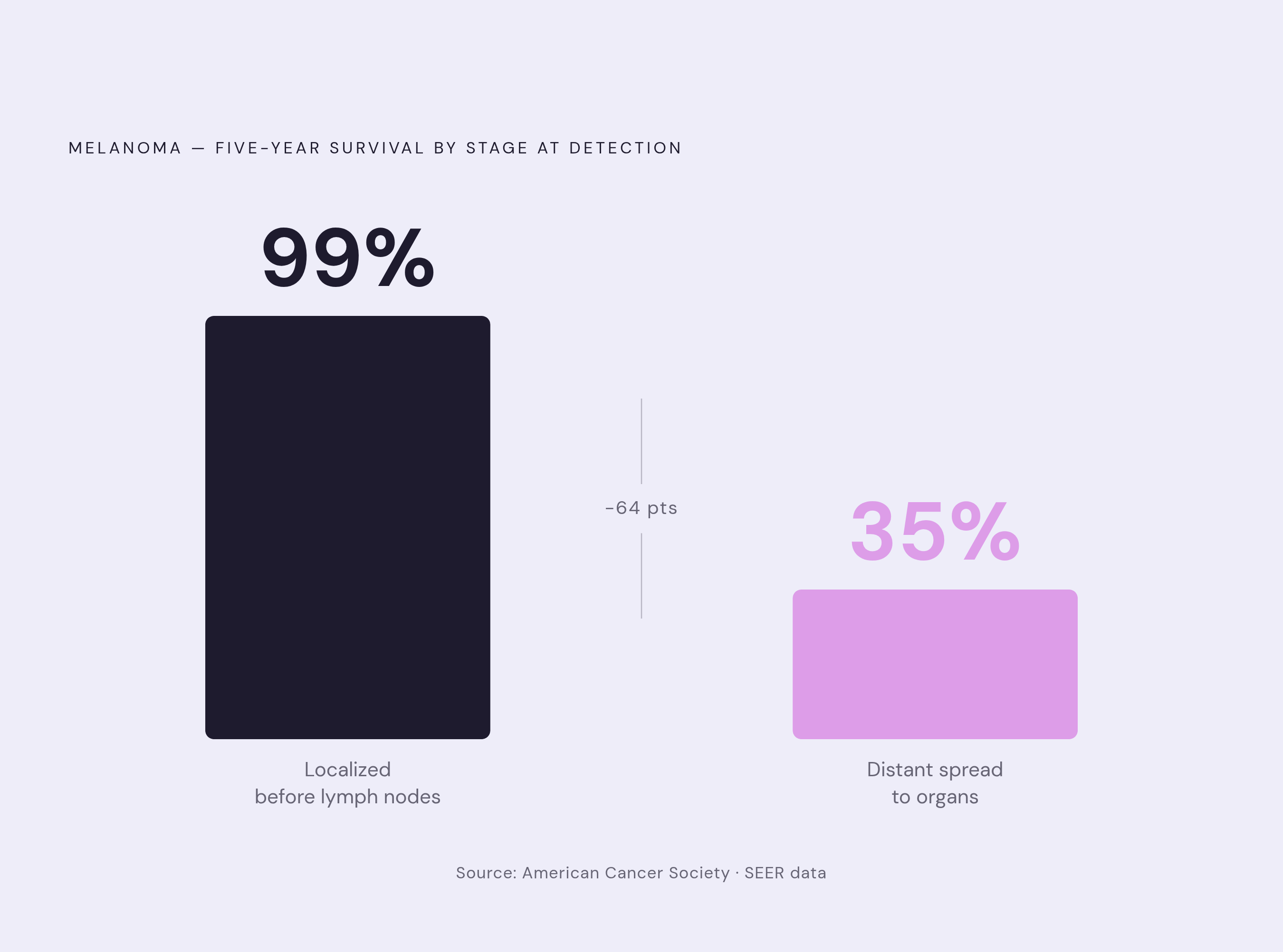
The survival data makes the argument for early recognition more compellingly than anything else. Melanoma caught before it reaches the lymph nodes carries a 99% five-year survival rate. Once it spreads to distant organs, that drops to 35%. The gap between those two numbers is what early recognition does in practice.
Visually, melanoma often presents as a new dark spot or a mole that is changing. Not necessarily large. Not necessarily dramatic. The early warning signs cluster around observable characteristics: asymmetrical shape, irregular or notched borders, multiple shades within a single lesion spanning tan through black, a diameter larger than a pencil eraser, and any evolution in size, shape, or new symptoms like itching or bleeding.
One form worth knowing specifically is amelanotic melanoma, which appears pink, red, or flesh-colored rather than dark. It's more aggressive than its pigmented counterpart and doesn't fit the expected mental image of a dangerous mole. Which is, of course, precisely what makes it dangerous.
Unlike BCC and SCC, melanoma can appear anywhere on the body, including areas with no meaningful sun exposure history. It can develop from a pre-existing mole or emerge as an entirely new lesion on previously clear skin. The geographic unpredictability is why full-body self-examination matters more than spot-checking familiar areas.
The ABCDE Rule and the Ugly Duckling Sign as Practical Self-Examination Tools
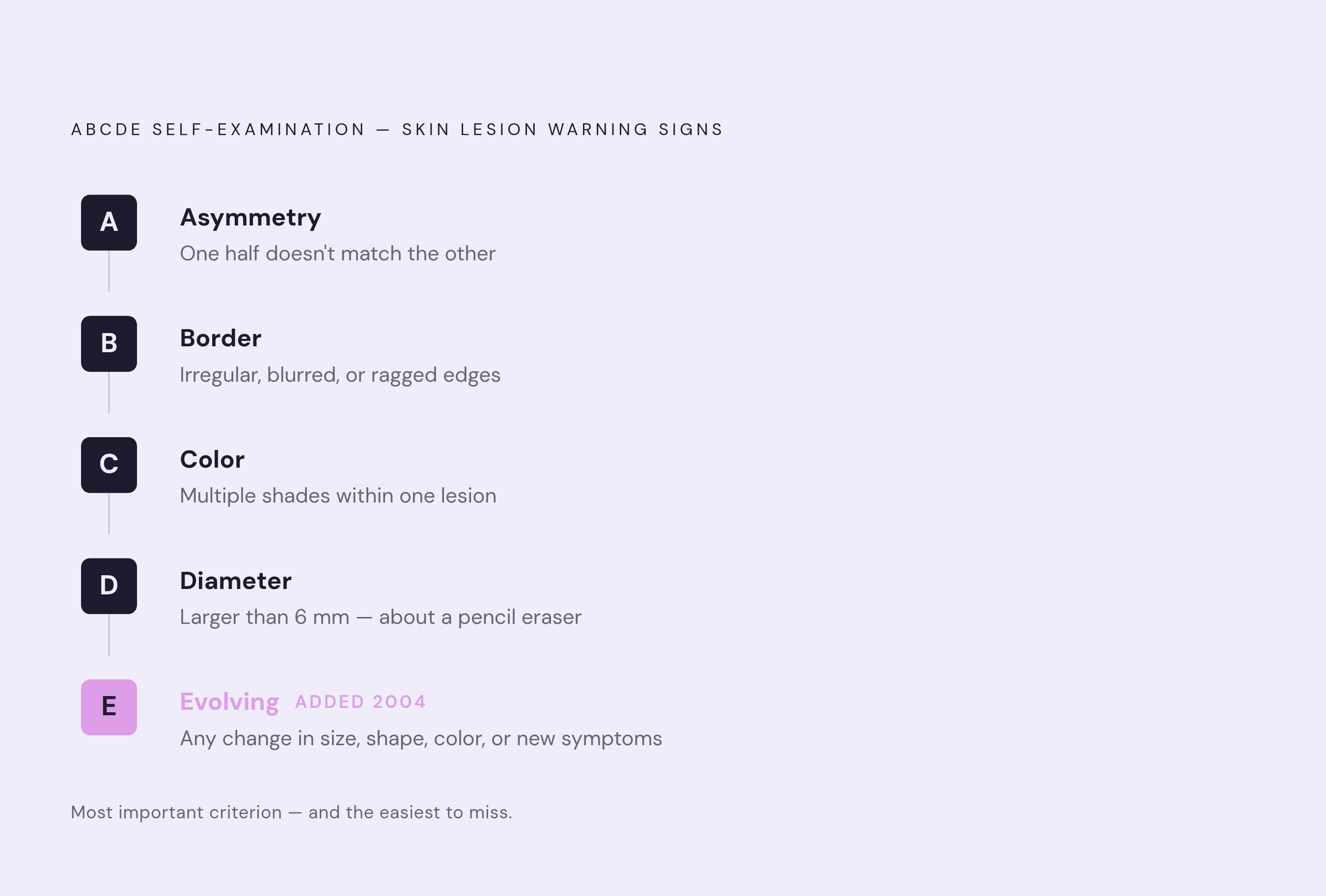
The ABCDE framework was introduced in 1985 to structure what clinicians were already observing in melanomas. Asymmetry, Border, Color, Diameter were the original four; Evolving was added in 2004. That fifth criterion is the most important one, and also the easiest to miss if you're not examining yourself systematically. A photograph captures a moment. Change is what slips past you.
To walk through each: Asymmetry means one half of a spot doesn't match the other if you drew a line through the middle. Border means the edges are irregular, blurred, or ragged rather than smooth. Color means multiple shades within a single lesion, or uneven pigment distribution. Diameter means the spot is larger than roughly 6mm (about the size of a pencil eraser), though size alone is not definitive. Evolving means anything has changed, whether shape, size, color, or new symptoms like itching, bleeding, or crusting.
The ugly duckling sign complements ABCDE without duplicating it. Most moles on a given person share a general family resemblance in size, color, and shape. A lesion that looks notably different from the rest (the outlier that simply doesn't fit the pattern) warrants attention regardless of whether it clears the ABCDE threshold. These two approaches catch different things, and they work best used together.
Some signs don't map cleanly onto either framework but still matter: a sore that doesn't heal within about four weeks, or a spot that repeatedly itches, bleeds, or crusts. These behavioral patterns, observed over time rather than seen in a single glance, are often more informative than how a lesion looks on any given day.
The mechanics of self-examination are worth being specific about. A monthly full-body check in good light, including the scalp, soles of the feet, between the toes, and under nails. Dated photographs of specific spots, because memory alone is unreliable for tracking subtle changes over weeks and months. Research supports the habit: patients who perform routine self-examination have better outcomes than those who don't, among people who go on to develop melanoma.
Self-examination is a trigger for evaluation, not a substitute for it. Worth stating plainly, because the line gets blurred.
Who Carries Higher Risk and Which Factors Compound Each Other
Fair skin, light eyes, and light hair reduce the protective effect of melanin, and that association dominates the popular imagination around skin cancer risk. But skin cancer occurs across all skin tones. Darker skin doesn't mean no risk; it means a different pattern of risk, often with lesions appearing in less sun-exposed areas and, consequently, being recognized later, sometimes at more advanced stages.
Severe sunburns before age 25 compound risk across all three major types. A personal or family history of skin cancer matters. A high number of moles, or the presence of atypical moles, matters. Immunosuppression, whether from medication or underlying condition, reduces the body's capacity to surveil and eliminate abnormal cells. Occupational or recreational sun exposure accumulated over decades matters. A history of tanning bed use matters.
The compounding dynamic is the part worth sitting with, though. A person with fair skin, multiple atypical moles, a history of childhood sunburns, and years of outdoor work isn't simply in a "higher risk" category. These factors don't just add; they interact and reinforce each other in ways no checklist fully captures. Most adults benefit from an annual professional skin examination, and the case for it strengthens with each additional overlapping factor.
What to Do When Something Looks Wrong, and Why Sooner Matters More Than Certainty
The threshold for getting something checked should be considerably lower than most people set it. A spot that is growing, bleeding, changing, or simply new and unfamiliar is sufficient reason to seek evaluation. You don't need to be certain something is wrong. Certainty is the clinician's job, not yours.
Waiting to be sure is the wrong frame entirely. The purpose of noticing a warning sign is to hand it off to someone with the tools and training to assess it, not to resolve it through extended observation at home.
When you do seek evaluation, documenting what you see before the appointment makes the conversation more useful. Dated photographs, notes on when the spot appeared, a description of how it has changed, its approximate size relative to a familiar object, its color and texture, whether it bleeds or itches: these details matter and are harder to reconstruct accurately after the fact.
Options for initial evaluation have expanded in useful ways. Store-and-forward teledermatology (in which a clinician reviews photographs without requiring an in-person visit) provides a lower-barrier first step for a new or changing spot, particularly when access to a dermatologist involves a long wait or significant travel. This isn't a replacement for in-person biopsy when one is warranted; it's often the right first move for triage. Nolla, an AI-assisted skincare telehealth service, uses AI-guided symptom intake to connect patients with licensed US clinicians who can review concerns and issue prescriptions in as little as 10 minutes, providing a faster path from initial observation to clinical review.
Annual professional skin exams remain the complement to monthly self-checks. Clinicians can evaluate areas that are difficult to self-examine, apply diagnostic tools unavailable at home, and assess ambiguous lesions with the full context of your history.
These cancers develop gradually, and early warning signs exist precisely because there is a detectable window before the disease advances. The goal isn't to become your own dermatologist. It's to become the kind of observer who knows when to make the call.