Types of Acne Lesions and What They Indicate
Microcomedones start every acne lesion, but inflammation and bacteria determine which type appears.

Every acne lesion originates in the same place: the pilosebaceous unit, the hair follicle and its attached sebaceous gland, sitting together in the dermis. Sebaceous glands produce sebum to lubricate the skin. That part is normal, even necessary. The problem is what happens when the process breaks down.
Androgens drive sebaceous gland activity. When androgen signaling rises, sebum production follows. But excess sebum is only one piece of it. The second is keratinization, the process by which cells inside the follicular canal shed and turn over. When those cells stick together instead of clearing out, they accumulate. More sebum plus stickier cells equals the conditions for a plug.
That plug, in its earliest form, is called a microcomedo. A hyperkeratotic cluster of cells forming in the lower follicular canal, invisible and asymptomatic. You cannot see it, feel it, or identify it by any conventional means. And yet it is the universal precursor to every acne lesion that follows. Before a whitehead, there was a microcomedo. Before a cyst, there was a microcomedo. The entire visible spectrum of acne begins with something you will not observe in a mirror.
What happens next depends on the follicle's local environment: whether the orifice is open or closed, how the immune system responds, which microbial strains are dominant.
This is where Cutibacterium acnes enters the picture, and where a common assumption falls apart. C. acnes is present on virtually all human skin, concentrated in sebum-rich areas. Most people who carry it do not develop significant acne. That observation should give anyone pause. Current evidence suggests the issue isn't the presence of C. acnes so much as strain-level imbalances and broader microbial ecosystem disruption. Viewed this way, acne looks less like a bacterial infection and more like a dysregulation problem, and that distinction shapes everything about how treatment gets approached.
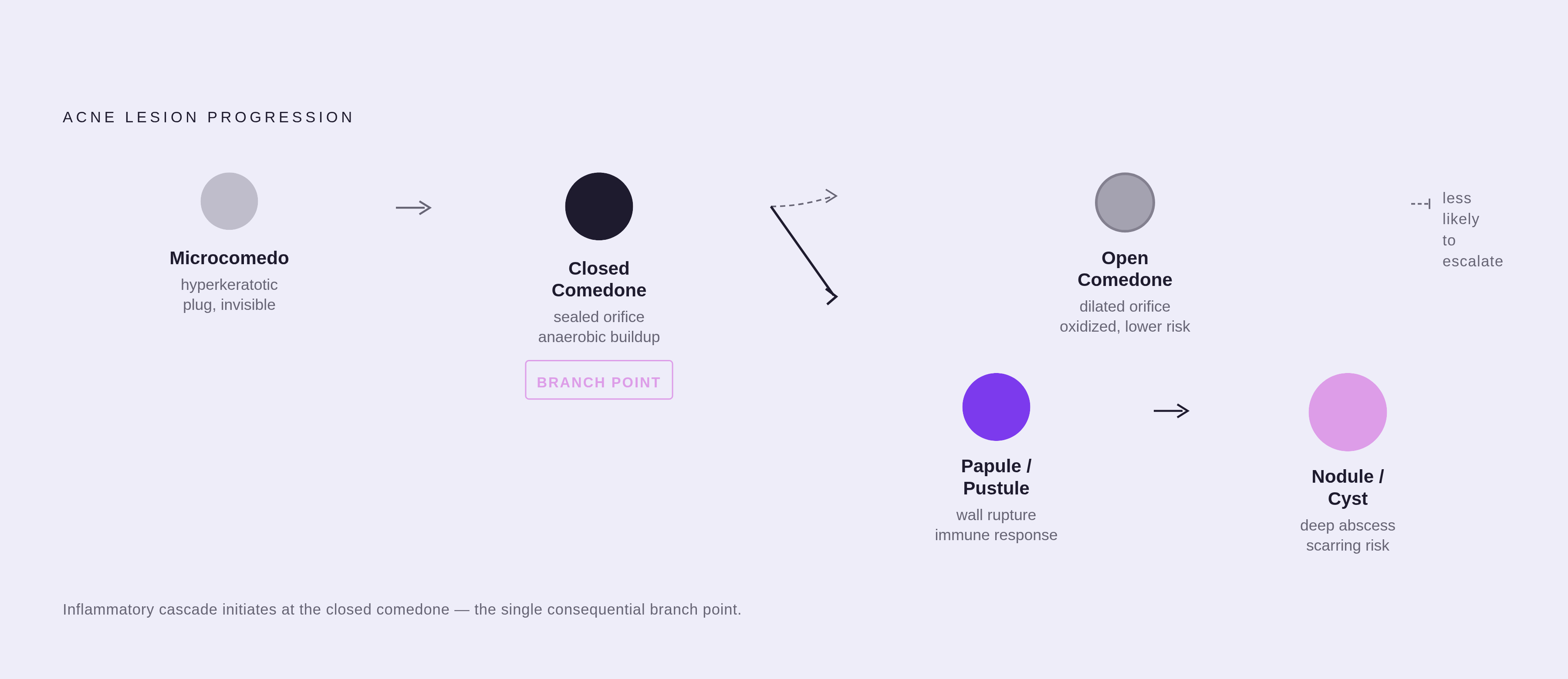
From the microcomedo, two paths diverge: comedonal lesions, where the immune system hasn't been recruited yet, and inflammatory lesions, where it has.
Closed and Open Comedones: What Blocked Follicles Look Like Before Inflammation Arrives
Blackheads and whiteheads are acne before the immune system gets involved. No inflammatory cascade is underway. The problem, at this stage, is almost purely mechanical: a blocked pipe.
Closed comedones form when the follicular orifice stays sealed. Keratin and sebum accumulate below the skin surface with nowhere to go, producing a small, dome-shaped bump, typically skin-colored or slightly grayish, smooth to the touch, without any surrounding redness. Easy to overlook, which is part of what makes them clinically interesting.
Here's something most people don't appreciate about closed comedones: they are the direct precursor to inflammatory acne. The sealed orifice means pressure builds inside an anaerobic environment where C. acnes proliferates unchecked. Treating them early is, functionally, treating acne before it escalates into something more damaging. A barely-visible bump is worth more attention than it typically gets.
Open comedones have a dilated orifice. Follicular contents are exposed to air, which causes oxidation and the characteristic dark color. Not dirt. Oxidized follicular material. This distinction matters because one of the most persistent instincts people have around blackheads is to scrub harder, which irritates the follicular wall in ways that actually invite the inflammation everyone is trying to avoid.
Because the orifice is open, some contents can extrude naturally. That partial pressure release makes open comedones less likely to progress to inflammatory lesions than closed ones. Still a keratinization and sebum problem; just a lower-urgency version of it.
What the comedonal category tells you, broadly, is that the skin's primary malfunction is happening upstream of the immune system. Bacteria haven't become the central story yet.
Papules and Pustules: When the Follicle Wall Fails and Inflammation Begins
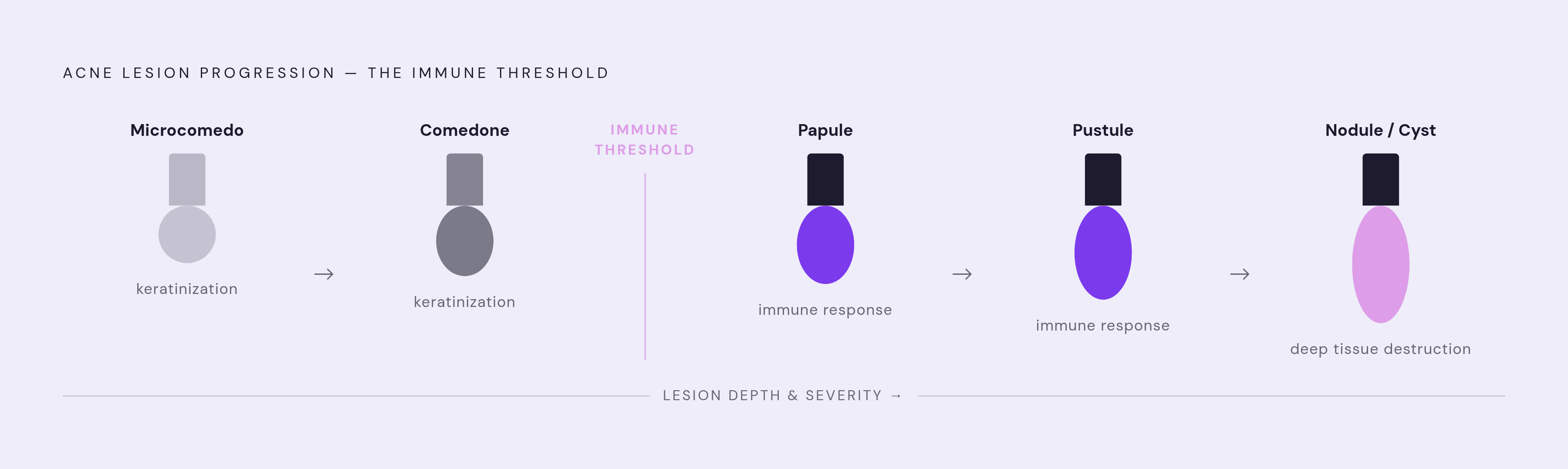
The transition from comedone to papule is where the biology changes categories entirely, not just escalates within one.
C. acnes, proliferating in the oxygen-poor environment of a closed comedone, metabolizes sebum and releases free fatty acids as a byproduct. Those free fatty acids irritate the follicular epithelium. The irritation signals the immune system. Neutrophils and lymphocytes are recruited, pressure inside the follicle rises, and eventually the follicle wall ruptures. Keratin, sebum, and bacteria spill into the surrounding dermis.
What forms at the surface is a papule: solid, raised, red, tender, typically less than a centimeter across, no visible head, no pus. The redness isn't incidental; it is the defining sign that inflammation has arrived. This is no longer a plumbing problem.
If the inflammatory response intensifies, neutrophils accumulate in sufficient concentration to form visible pus, and the papule becomes a pustule: a raised lesion with a whitish or yellowish head, surrounded by inflamed skin. The pus is a concentration of immune cells, not evidence of infection spreading systemically. The visible head marks the point where the lesion has reached the upper dermis or skin surface.
Both papules and pustules typically resolve within two weeks, assuming nothing is done to worsen the situation. The skin, at this tier, can recover without permanent marks if the follicle isn't manually disrupted. That caveat becomes significant when we get to scarring.
Treatment priorities shift here in ways that matter. Pore obstruction is still part of the picture, but bacterial activity and the inflammatory cascade are now central. An approach targeting only keratinization is addressing part of the problem while leaving the more urgent part unaddressed.
Nodules and Cysts: Deep Tissue Involvement and Why These Lesions Behave Differently
Everything discussed so far involves, in varying degrees, the upper layers of the skin. Nodules and cysts operate in a different anatomical register, and that difference changes the clinical equation substantially.
A nodule is a solid, hard, painful lump with no visible head. The inflammation is extensive but contained within tissue. Nodules can persist for months rather than the days-to-weeks timeline of papules and pustules, and during that persistence, the inflammatory process is continuously degrading surrounding tissue.
A cyst takes the response further: large, fluid-filled, painful, soft to the touch because pus has accumulated deep within. The follicle has, functionally, become an abscess. When nodules and cysts occur together, the presentation is called nodulocystic acne, one of the most severe expressions of acne vulgaris.
Two things follow directly from the depth of these lesions. First, topical treatments cannot reach the dermis where nodules and cysts originate. Applying benzoyl peroxide or a retinol serum over a nodule isn't just insufficient; it's physiologically mismatched to the problem. The active ingredient never arrives where the problem is. Second, manipulation creates a specific mechanical hazard: inflammatory contents under high pressure in deep tissue, when squeezed, are forced laterally through surrounding dermis rather than upward and out. That lateral spread extends inflammatory damage through tissue that would otherwise recover. The risk of permanent scarring is not theoretical here; it follows directly from the physics of what happens when you press on a sealed, pressurized pocket in deep tissue.
The point isn't "these are worse pimples." The biology has escalated to a point where the therapeutic requirements are categorically different from what works at the comedonal or papular stage. It is a different problem.
How Lesion Type Maps to Acne Severity Grading
When a clinician assesses acne severity, the process is more structured than a general impression. They are counting lesions by category: non-inflammatory (comedones), inflammatory (papules and pustules), and nodulocystic (nodules and cysts). Those categories map onto a grading framework that determines which treatments are clinically appropriate.
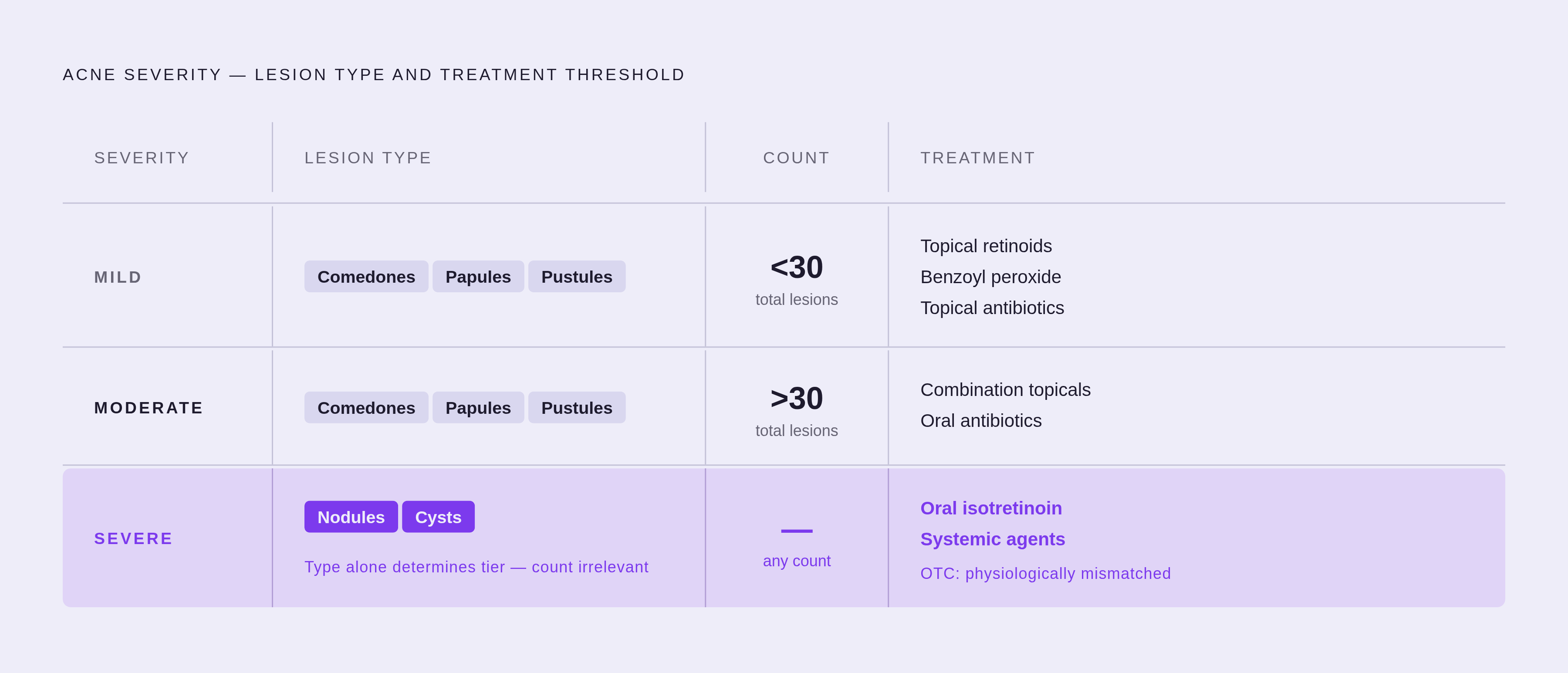
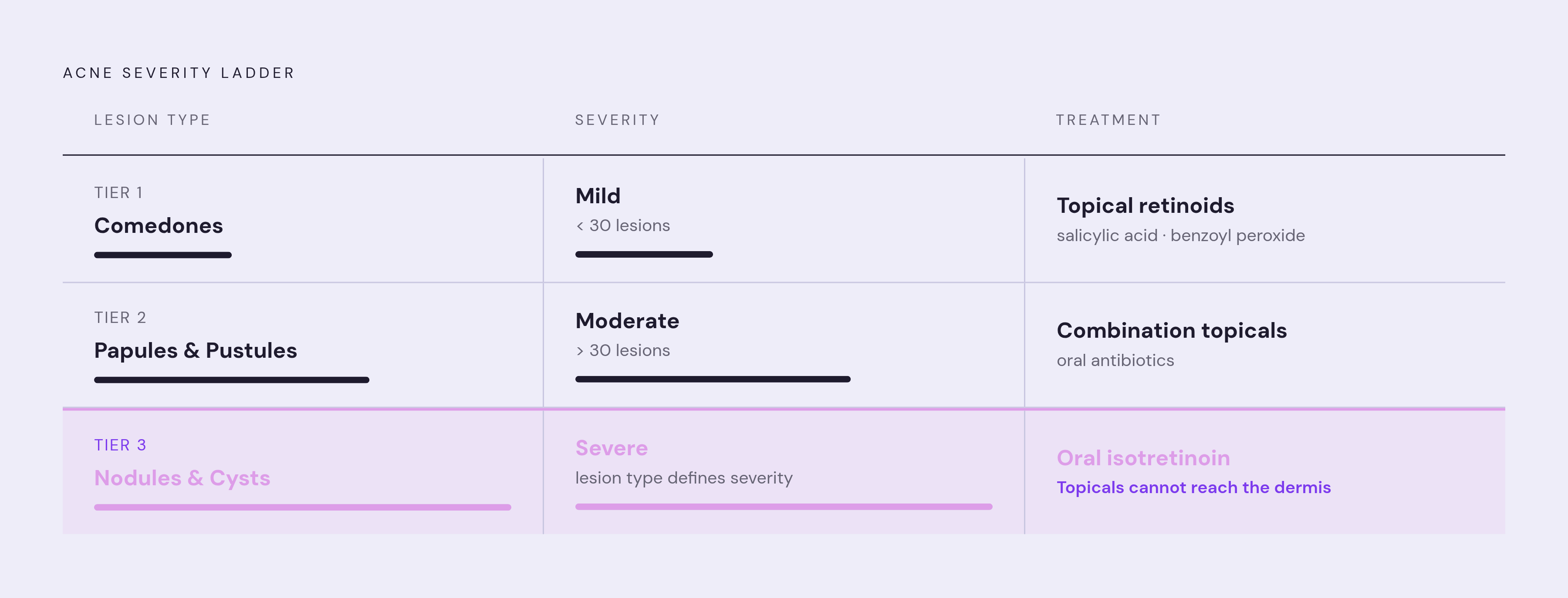
Mild acne is characterized primarily by comedones with a limited number of papules and pustules, generally a total lesion count under 30. Moderate acne adds more papules and pustules to the comedonal base, typically above 30. Severe acne is defined largely by the presence of nodules and cysts, at which point the specific count matters less than the lesion type itself.
Lesion count alone, though, is not the full picture. Severity grading also accounts for whether scarring is already present, the psychological burden, whether draining lesions or sinus tracts have formed, and how a patient has responded to prior treatments. A single cyst on an otherwise clear face in someone with significant scarring from previous cycles represents a different clinical reality than a high comedone count with no scarring history.
This framework has practical consequences because it governs what belongs on the therapeutic menu. At the mild end: topical retinoids, benzoyl peroxide, topical antibiotics. At the moderate tier: combination topicals, possibly oral antibiotics. At the severe end, where nodulocystic lesions are present, systemic agents like oral isotretinoin become relevant. Over-the-counter approaches aren't merely insufficient at this tier; they are mismatched to the biology.
Why Some Acne Leaves Marks and Others Don't: Lesion Type as a Predictor of Scarring and PIH
Retrospective data makes the relationship between lesion type and lasting marks fairly clear. Comedonal acne is significantly more prevalent among patients who develop neither scarring nor post-inflammatory hyperpigmentation. Nodular acne is significantly more prevalent among patients who develop both. Lesion type functions as a predictor, not just a descriptor.
Two distinct categories of marks emerge from acne, and they have different origins.
Post-inflammatory hyperpigmentation, PIH, is the dark discoloration that remains after a lesion resolves. It is not a scar. It is the skin's melanin response to inflammation, and its prevalence skews substantially by skin phototype. In one study, PIH prevalence reached 65% in African American patients, 48% in Hispanic patients, and 25% in Caucasian patients. Darker phototypes carry meaningfully higher risk not because the acne is more severe but because the melanocytes respond more vigorously to inflammatory stimuli. A large international survey published in JAAD found that 71% of people with PIH had previously experienced acne, and 85% believed earlier treatment could have prevented it. In the majority of cases, PIH takes more than a year to fade on its own.
Physical scarring, the pitted or keloidal marks, is a different phenomenon driven by deep tissue destruction. When the dermis is damaged repeatedly or severely, the collagen architecture doesn't fully reconstitute. Nodules and cysts, persisting for weeks or months in deep tissue, create exactly those conditions. Manipulation accelerates damage by forcing inflammatory contents laterally, extending injury beyond the lesion boundary.
The practical consequence: correctly identifying a lesion, specifically recognizing when something is a nodule rather than a large pustule, changes the urgency of the decision you make about it. Leaving it alone and seeking appropriate treatment sooner is directly connected to whether permanent marks result.
Triggers That Shift the Lesion Spectrum Toward More Inflammatory Types
Triggers don't affect all lesion types equally. Some drive excess sebum production, feeding comedone formation at the upstream end of the process. Others amplify the inflammatory response, pushing progression toward deeper, more damaging lesions. Knowing which category a trigger belongs to adds precision to the question of why a particular flare is happening.
Hormonal drivers operate upstream. Androgens stimulate sebaceous gland growth and increase sebum secretion; this is the foundational mechanism behind comedone formation. Sebaceous glands in the lower face carry a higher density of androgen receptors than other facial zones, which explains why hormonal flares characteristically concentrate along the jaw and chin. Conditions that elevate the androgen-to-estrogen ratio, including polycystic ovary syndrome and insulin resistance, compound the upstream sebum problem. Insulin resistance also affects IGF-1, a growth factor that increases both sebum production and keratinocyte proliferation, simultaneously. Persistent lower-face breakouts that don't respond to conventional topicals are worth discussing with a clinician, not just treating with a stronger product.
Dietary triggers work through related hormonal pathways. High-glycemic foods provoke an insulin spike, which elevates IGF-1, which increases sebum production and androgen synthesis simultaneously. Dairy, particularly skim milk, has been associated with acne flares in a portion of patients; the proposed mechanism involves IGF-1 and hormones present in milk from pregnant cows. The evidence is associative rather than conclusive, but the biological plausibility is substantial enough to take seriously.
Stress operates through cortisol and corticotropin-releasing hormone, both of which stimulate sebaceous gland activity directly. Chronic stress doesn't produce a single hormonal perturbation; it disrupts sleep and dietary patterns as well, compounding the pathways that push toward more sebum and more inflammation.
For someone observing their own skin, the types of lesions appearing during a flare offer feedback on which biological pathway is most active. Predominantly comedonal breakouts suggest a sebum and keratinization driver. A sudden shift toward papules, pustules, or cysts suggests inflammation is being amplified beyond simple pore obstruction. Recognizing which is happening first clarifies which lever is actually worth addressing.
What Lesion Type Actually Tells You About What Your Skin Needs Next
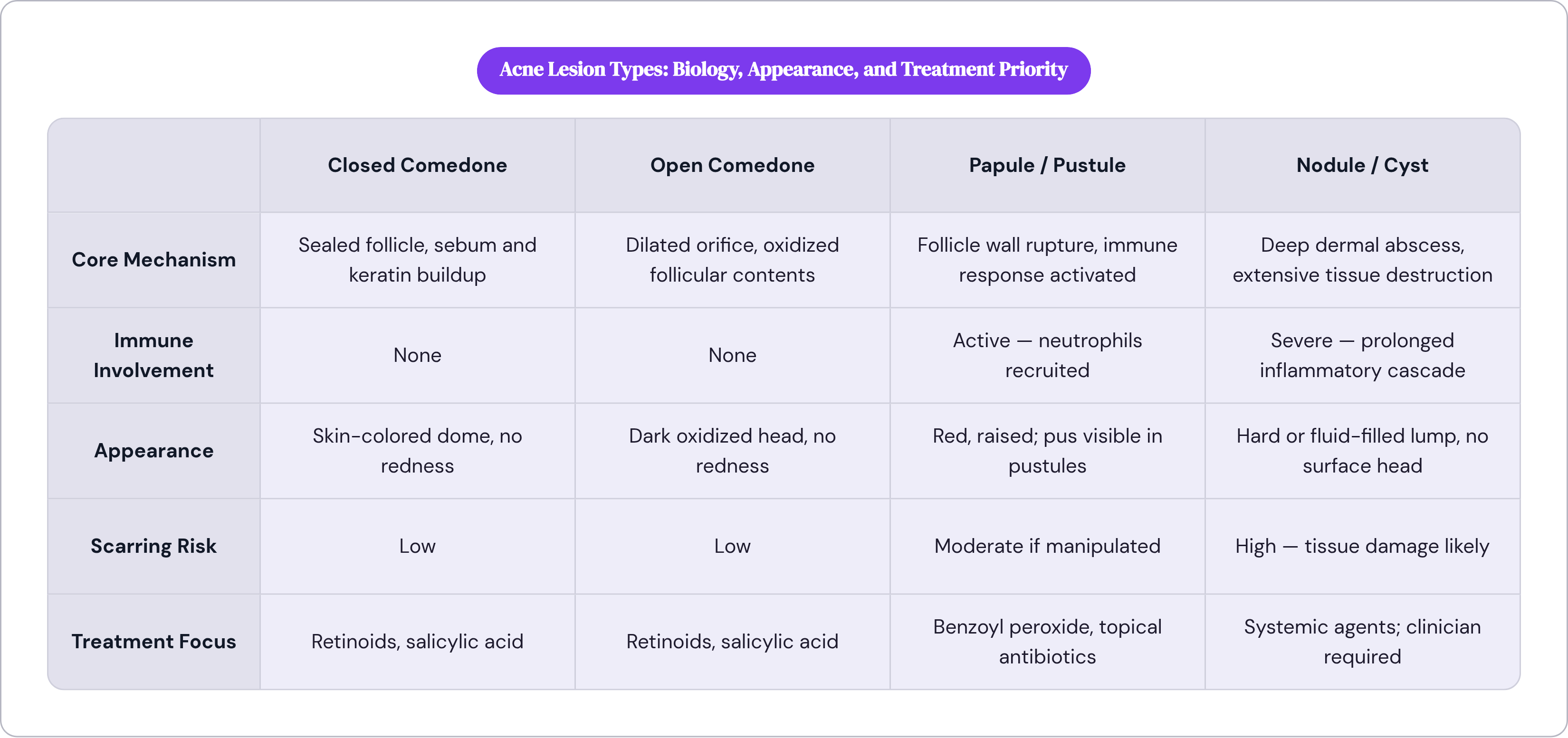
Lesion type is diagnostic information. Not a cosmetic taxonomy, not a severity scale to memorize. Each category points toward a different biological mechanism and, therefore, a different category of intervention.
Comedones are a keratinization and sebum story. Topical retinoids work here because they normalize keratinization at the follicular level; salicylic acid works because it dissolves the keratin plugs forming inside the pore. Consistency matters more than intensity. Because microcomedones are invisible and constantly forming, treatment has to be sustained to stay ahead of the process. A weekend application schedule won't hold the line.
Papules and pustules require a broader target. Bacterial activity and immune response are now central, which is where benzoyl peroxide and topical antibiotics enter. Benzoyl peroxide is particularly useful because C. acnes has not demonstrated meaningful resistance to it, unlike conventional antibiotics. Manual manipulation actively worsens things at this stage, not for aesthetic reasons but mechanical ones: squeezing drives the inflammatory cascade deeper, extends the lesion's timeline, and substantially raises the probability of PIH.
Nodules and cysts are where the over-the-counter aisle stops being relevant, full stop. This isn't a matter of finding a stronger product or applying it more diligently. Topical actives cannot reach the dermis where these lesions originate. Clinician involvement at this tier isn't a recommendation; it's a practical necessity dictated by anatomy. Waiting to see if a nodule resolves on its own risks months of cumulative tissue damage and a meaningfully higher probability of permanent marks, because it often does eventually resolve, just not without cost.
There's a version of skincare literacy that treats all breakouts as points on a single spectrum, varying in intensity but not in kind. The biology doesn't support that. A whitehead and a cyst emerge from the same upstream origin but represent distinct disease states by the time they're visible. Getting that identification right, even roughly, matters in ways that go beyond knowing which aisle to shop in.