Epidermis Layers and Their Protective Roles
The deepest skin layer generates new cells, produces melanin for UV protection, and detects touch.

The stratum basale is the deepest layer of the epidermis, a single cell thick, pressed against the dermis below. This is where keratinocytes originate through continuous cell division. Every new skin cell begins here. No other source exists.
But the basale is not only a keratinocyte factory. Scattered throughout this layer are melanocytes, the cells responsible for producing melanin. Melanin comes in two forms: eumelanin, which produces darker pigmentation, and pheomelanin, which produces pink and red tones. Both filter ultraviolet radiation and protect the DNA of living cells from UV-induced damage. Your body's internal sunscreen begins here, not at the surface, but at the deepest living layer of the skin.
Merkel cells also reside in the basale. These are mechanoreceptors, tuned to detect light touch. So the basale is doing three distinct things at once: generating new cells to feed the layers above, initiating UV protection from within, and relaying sensory information about physical contact. Proliferative, photobiological, and neurological work, running in parallel.
Why does this matter? Because the basale is the only source of new keratinocytes. If it is damaged by radiation, chronic inflammation, or disease, the entire regenerative pipeline above it is compromised. Nothing upstream can compensate. The roughly 30-day timeline it takes for a keratinocyte to complete the full journey from basale to surface depends on healthy, consistent cell division here. When that falters, the consequences travel upward through every layer.
The transit through the stratum spinosum and stratum granulosum: where cells transform into barrier material
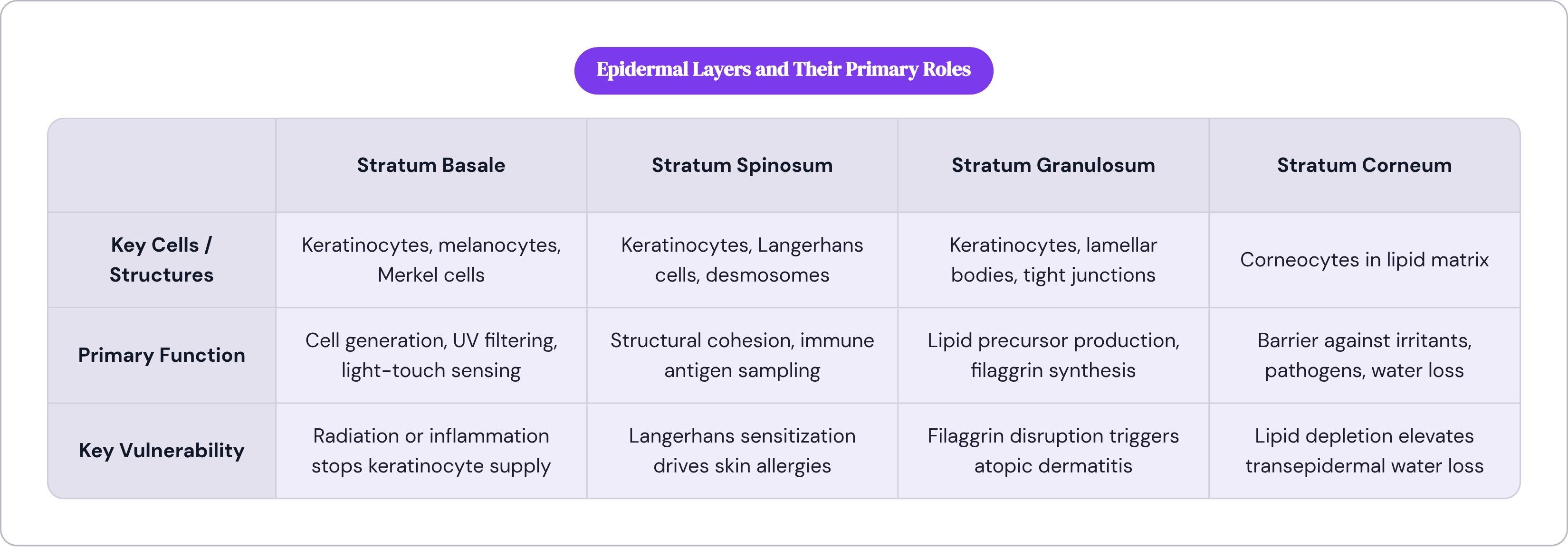
Once a keratinocyte leaves the basale, it enters the stratum spinosum, named for the spiny projections visible under a microscope. Those projections are desmosomes, protein structures that connect adjacent cells and give this layer its mechanical cohesion. The cells are beginning to flatten, and they are beginning to synthesize the structural proteins they will carry upward.
The spinous layer is where Langerhans cells live, and they are worth understanding clearly. These are the epidermis's resident immune sentinels, capable of detecting foreign substances, sampling antigens, and coordinating immune responses. Their placement here is not incidental. It positions immune detection close to the surface while maintaining a buffer of living tissue between the threat and the bloodstream. Here is the complication, though: Langerhans cells are also implicated in the development of skin allergies. The same system that guards you can sensitize you. Immune protection involves trade-offs, and that tension runs through almost every skin condition worth understanding.
Above the spinous layer, keratinocytes enter the stratum granulosum. The name comes from granules that become visible as cells begin programmed death. These granules contain lipid precursors packaged in structures called lamellar bodies. As cells move through this layer, those precursors are extruded into the intercellular space above, where they assemble into the lipid matrix that gives the stratum corneum its barrier function. Ceramides, cholesterol, and free fatty acids are all produced here before being deposited above.
The granular layer is also where filaggrin is synthesized. Filaggrin aggregates keratin filaments and is essential for the structural integrity of the flattened, protein-dense cells that will populate the stratum corneum. When filaggrin expression is disrupted, the consequences are specific and clinically significant, and the most important one involves atopic dermatitis.
Tight junctions also form in the granular layer. These protein complexes regulate what passes upward and establish distinct pH zones within the stratum corneum above. That pH regulation is not trivial.
By the time a cell exits the granular layer, it is no longer living. But this is not passive deterioration. It is a programmed differentiation into barrier material. Everything that made it a metabolically active cell has been metabolized into something structurally useful. The underlying logic of the epidermis is that life produces barrier.
The stratum lucidum: a transitional layer present only in thick skin
The stratum lucidum is thin, translucent, and composed of dead, flattened keratinocytes. It appears only in the thick, hairless skin of the palms and soles of the feet. In most of the skin on your body, it is simply absent.
Its function is mechanical. In areas subject to high friction and compressive force, it provides an extra transitional zone between the granular and corneum layers, adding cushioning where abrasion is greatest. It is an anatomical adaptation to mechanical stress, not a universal architectural feature.
For anyone following the five-layer count and finding the lucidum missing in most locations, the absence is not a gap in the system. The stratum corneum forms properly in thin-skinned areas regardless. The lucidum is an evolutionary refinement for specific conditions of load-bearing and friction, and the fact that it exists at all is a useful reminder that the epidermis is not a generic structure. It adapts its architecture to the demands of the region it covers.
What the stratum corneum actually does: and why it is more than a dead outer shell
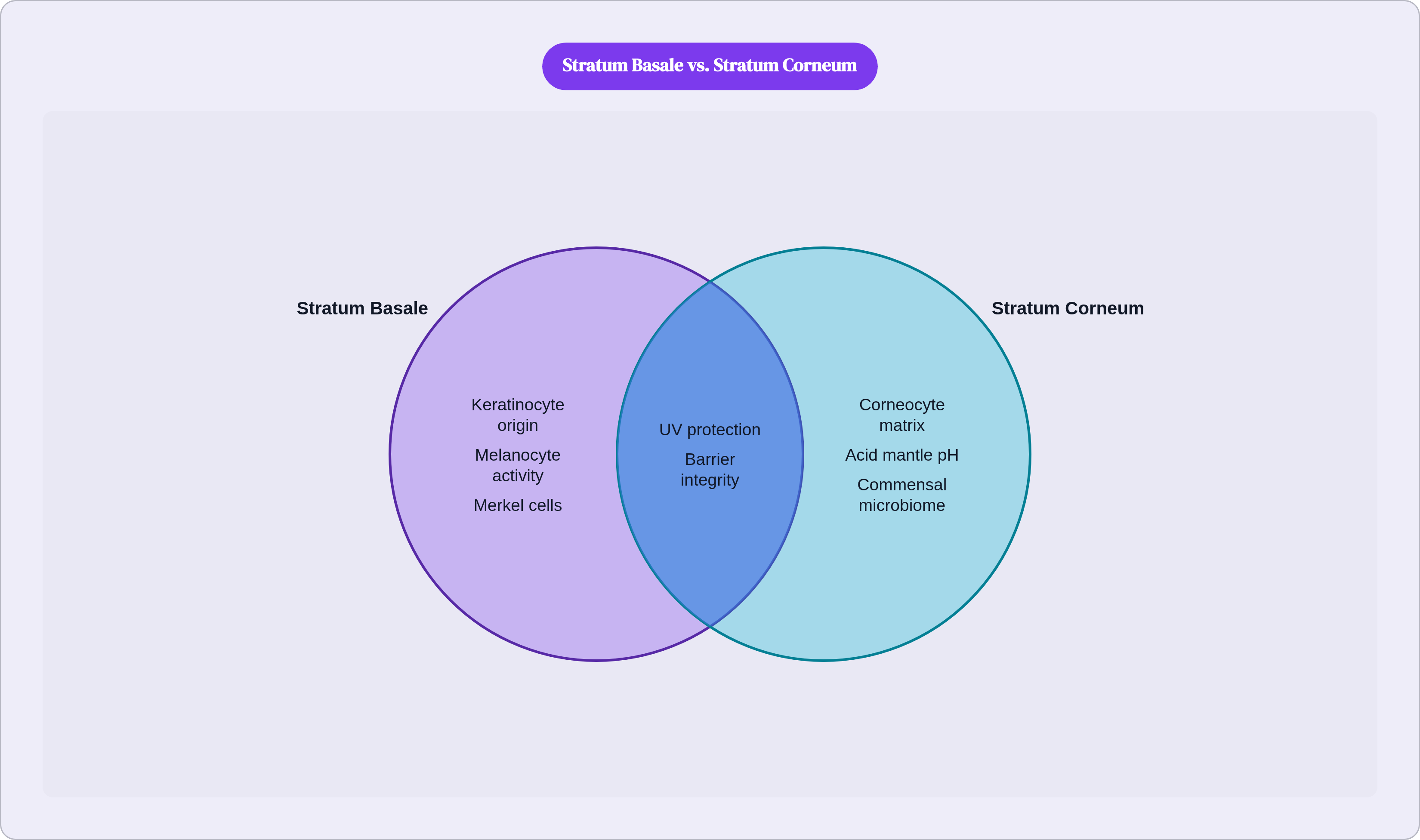
After roughly 30 days of differentiation, migration, and transformation, a keratinocyte arrives at the stratum corneum. By this point it has surrendered its nucleus, flattened into a protein-dense disk called a corneocyte, and taken its place in a lipid matrix assembled from the precursors produced in the granular layer. The Cleveland Clinic puts daily cell shedding at approximately 40,000 skin cells. As new cells complete the cycle below, the oldest corneocytes at the surface are continuously released.
The architectural description that has become standard in dermatology is "brick and mortar": corneocytes as bricks, the surrounding lipid matrix as mortar. I find it useful not because it is elegant but because it is accurate. The lipid mortar is a precise mixture of ceramides, cholesterol, and free fatty acids, and none of those three components is interchangeable with the others. Ceramides alone constitute approximately half of the lipids in the stratum corneum, which is why ceramide depletion compromises barrier function so disproportionately. But replacing ceramides without cholesterol and free fatty acids leaves the lamellar structure incomplete. The mortar only works when all three are present in roughly the right proportions.
What this barrier actually does is worth spending a moment on, because people tend to think of it in one direction only. The stratum corneum controls inward and outward movement simultaneously. It resists the inward passage of irritants, allergens, and pathogens. It prevents transepidermal water loss outward, keeping the body's hydration from evaporating through the skin. Both directions matter equally. Thinking of the stratum corneum as primarily a lock on the outside misses half of what it does.
There is also an antimicrobial function built into the surface chemistry. The acid mantle, with a surface pH of roughly 4.0 to 5.8, creates a chemical environment hostile to most pathogens. There is an antioxidant capacity that neutralizes free radicals before they can reach living cells below. And the photoprotective function here complements the melanin-based UV filtering that begins at the basale.
Recent imaging research has revealed something I find notably interesting: the pH within the stratum corneum is not uniform. There are three distinct zones, a moderately acidic lower zone, a more acidic middle, and a pH-neutral upper zone, each supporting different enzymatic and antimicrobial activity. The tight junctions formed in the granular layer help establish and maintain these zones. The stratum corneum is not a monolith. It is a graded environment, and we are only now beginning to map it at the resolution it deserves.
How the microbiome and immune system turn the epidermis into an active defense network
The stratum corneum is not sterile. It is colonized by a commensal microbiome, bacteria, fungi, and other microorganisms living on and within the outermost layers of skin. This microbiome is not an incidental occupant. It actively influences immune responses and metabolic processes in the tissue below it.
The acid mantle's role here is more selective than people realize. At a pH of 4 to 5.8, it does not simply suppress pathogens uniformly. It suppresses harmful bacteria while supporting the growth of beneficial commensal species. It is selective chemistry, and it depends on a structurally intact stratum corneum to maintain it. When the barrier is compromised, the mantle shifts. When the mantle shifts, the microbial community changes. That is not a metaphor for disruption; it is the actual mechanism.
Langerhans cells complete their role in this system by migrating to lymph nodes and initiating adaptive immune responses once they have sampled a relevant antigen. They are the link between what is detected at the epidermis and what the immune system does about it. Their position in the spinous layer, rather than at the surface, is strategic: they operate behind the first line of defense, not at it.
What I keep coming back to, having read through a fair amount of recent literature on this, is the framing of the epidermis as a "dynamic immunological interface." The phrase earns its complexity. Keratinocyte differentiation, lipid metabolism, tight junction integrity, hydration regulation, and innate immune signaling are coordinated functions; they are not independent systems that happen to coexist. A product or habit or condition that disrupts the stratum corneum does not simply dry out the skin. It shifts the microbiome composition, alters the pH gradient, and opens immunological vulnerabilities that begin at the surface and ripple downward. The disruption travels the same direction as the cells, just in the opposite direction from where it started.
What happens when the barrier breaks down: conditions that begin in the epidermis
Barrier dysfunction is the mechanism underlying several well-characterized skin conditions. Atopic dermatitis, psoriasis, adult acne, and ichthyosis all involve some failure at the level of epidermal architecture. These are not primarily cosmetic problems. They are failures of a system that is inseparable from immune function, hydration regulation, and infection resistance.
In atopic dermatitis, a central driver is the overactivation of an inflammatory signaling axis involving two cytokines, IL-13 and IL-4. This overactivation downregulates filaggrin expression in the granular layer. Less filaggrin means structurally compromised corneocytes, which means a compromised lipid matrix, which means elevated transepidermal water loss and increased permeability to allergens and pathogens. The line from molecular biology to clinical symptom is direct and traceable. What presents as dry, itchy, inflamed skin is a cascade that begins in the granular layer with a disruption to a single protein.
Psoriasis operates through a different mechanism but arrives at a similarly dysfunctional endpoint. Keratinocyte hyperproliferation compresses the normal 28-to-30-day cell cycle to as few as 3 to 10 days. Cells reach the stratum corneum before they have completed differentiation. They deposit immature, improperly assembled material in the place where a functional barrier should be. The characteristic scaling and inflammation reflects, at the cellular level, a stratum corneum populated with cells that never had time to become what they were supposed to become.
Both conditions produce elevated transepidermal water loss, microbial susceptibility, and amplified inflammatory signaling. These consequences map directly onto the barrier functions described earlier. When those functions fail, the symptoms follow predictably: persistent dryness, itching, discoloration, heightened sensitivity, recurring infections, spreading inflammation. They are not separate problems to be treated individually. They are manifestations of a single architectural failure.
How aging changes the epidermis and why barrier resilience declines over time
Aging is largely a story of slowing. The stratum basale divides more slowly, fewer new keratinocytes are generated per unit of time, and the 30-day renewal cycle lengthens. Every layer above depends on that supply of new cells from below, and when the supply diminishes, every layer above is affected.
Ceramide production declines with age. The lipid mortar thins. Permeability barrier function weakens, and transepidermal water loss increases. As one Cleveland Clinic dermatologist put it, the stratum corneum simply does not bounce back the way it once did after a day in the sun or after exposure to a harsh product. That is not vague. It reflects a concrete reduction in both the rate and quality of cell production in the layers below.
Melanocyte density also decreases with age, and this is significant in a way that does not get enough attention. The skin's endogenous UV filtering capacity is declining at the same time that cumulative sun exposure continues to mount. Older skin is not simply more fragile cosmetically. It is less capable of protecting its own DNA from the same level of UV radiation that younger skin would manage with greater reserve.
Langerhans cell density declines as well. Reduced immune surveillance in the spinous layer means slower detection of pathogens and antigens, and a diminished capacity to initiate an appropriate adaptive immune response. The epidermis becomes immunologically slower at the same time it becomes structurally thinner. Those two changes are not independent of each other.
The practical consequence is that behaviors and products that were tolerable in earlier decades become measurably damaging as the regenerative margin narrows. Hot water, aggressive exfoliation, skipping barrier-supportive ingredients: these are not merely suboptimal choices in older skin. They are choices made against a system that has measurably less capacity to recover from insult.
Ingredients and habits that support each layer's function: and the ones that undermine it
Supporting the epidermis from the outside is not a matter of adding more. It is a matter of working with the biology rather than against it.
The stratum corneum's lipid matrix is built from ceramides, cholesterol, and free fatty acids in a specific ratio. Formulations that include all three lipid components together outperform ceramide-only products in restoring barrier function because the lamellar structure of the lipid mortar requires the full complement to assemble correctly. Ceramides constitute roughly half of stratum corneum lipids, which explains why their loss is disproportionately damaging, but replacing ceramides alone leaves the mortar structurally incomplete.
Niacinamide, in concentrations between 4% and 10%, works differently and at a different level. It stimulates the skin's own production of ceramides and fatty acids and addresses the inflammatory component of barrier disruption that topical lipid replenishment alone cannot reach. It operates on the granular and spinous layers, not just the surface. That distinction matters: niacinamide is doing something that no amount of topically applied ceramide can replicate, because it targets the synthetic machinery rather than supplementing the output.
Retinoids accelerate keratinocyte turnover in the basal and spinous layers. The clinical evidence for their efficacy in acne treatment and skin aging is robust. Because they speed the cell cycle, they can transiently disrupt the stratum corneum during early use, as cells are pushed upward faster than the differentiation and lipid assembly processes can keep pace. Starting at a low concentration, building slowly, and consistently using broad-spectrum sunscreen are standard guidance for concrete reasons.
Panthenol, glycerin, hyaluronic acid, and squalane provide humectant hydration, anti-inflammatory support, and lipid compatibility. Their role is to improve the tolerability and effectiveness of the primary barrier repair actives. They are useful, but their function is supportive in the precise sense of the word; they are not substitutes for the actives themselves.
Some habits cause damage that is specific and mechanistically understood. Daily use of chemical exfoliants, or combining multiple exfoliating products, strips the lipid layer faster than it can regenerate. Sulfate-containing foaming cleansers and hot water disrupt the lipid structure of the stratum corneum directly. Aggressive use of powered cleansing devices disrupts corneocyte cohesion at a mechanical level. These are not theoretical risks. They are predictable outcomes of applying known stressors to a structure whose vulnerability has been characterized.
One important caveat: the evidence supports specific active ingredients, not every formulation that contains them. Concentration, delivery system, and individual biological variation all influence efficacy. Products are not interchangeable simply because they share an ingredient name on the label.
Sunscreen deserves its own mention. The melanocytes in the stratum basale provide real, meaningful endogenous photoprotective capacity, but that capacity is finite and decreases with age. Broad-spectrum sunscreen is the external complement to internal photoprotection. Its importance grows, not diminishes, as melanocyte density declines over time.
When skin symptoms suggest barrier failure that goes beyond what topical care can address
There is a point at which barrier disruption moves into territory that topical care cannot meaningfully address, and recognizing that threshold is as important as understanding the biology that precedes it.
Persistent symptoms are the signal. Recurring inflammation that does not resolve with consistent barrier support, infections that return after treatment, rashes that spread despite appropriate care: these patterns suggest that the disruption has moved into immunological territory. The epidermis is deeply integrated with the immune system, and when that integration fails at a systemic level, the treatment required is systemic as well.
Atopic dermatitis and psoriasis are clinical diagnoses. They require clinical evaluation and, in many cases, intervention beyond anything applied to the skin's surface. This is not a statement about the limitations of skincare in a general sense. It is a statement about mechanism. Filaggrin downregulation driven by IL-13 and IL-4 overactivation is not correctable by ceramide application. Keratinocyte hyperproliferation driven by immune dysregulation in psoriasis is not addressable by any topical ingredient currently available.
A randomized controlled trial examining guselkumab, a targeted anti-IL-23 therapy, in plaque psoriasis found that lesional stratum corneum barrier function and ceramide profiles normalized with treatment. The barrier was restored not by addressing the barrier directly, but by addressing the immune signaling that was preventing the barrier from forming correctly. That result carries a specific implication: the stratum corneum we can see and touch is, in conditions like these, a downstream indicator of something happening much further upstream in the biology. Getting that upstream process right requires a clinician, a diagnosis, and often a systemic intervention.
Skincare is not medicine, and medicine is not skincare. Knowing which problem you are dealing with depends on knowing how the system is supposed to work, and at what point it has exceeded the capacity of topical support to repair. Understanding the layers is what makes that distinction possible.