Skin Cancer Symptoms on the Face by Type
Recognize which facial spot signals which cancer type.
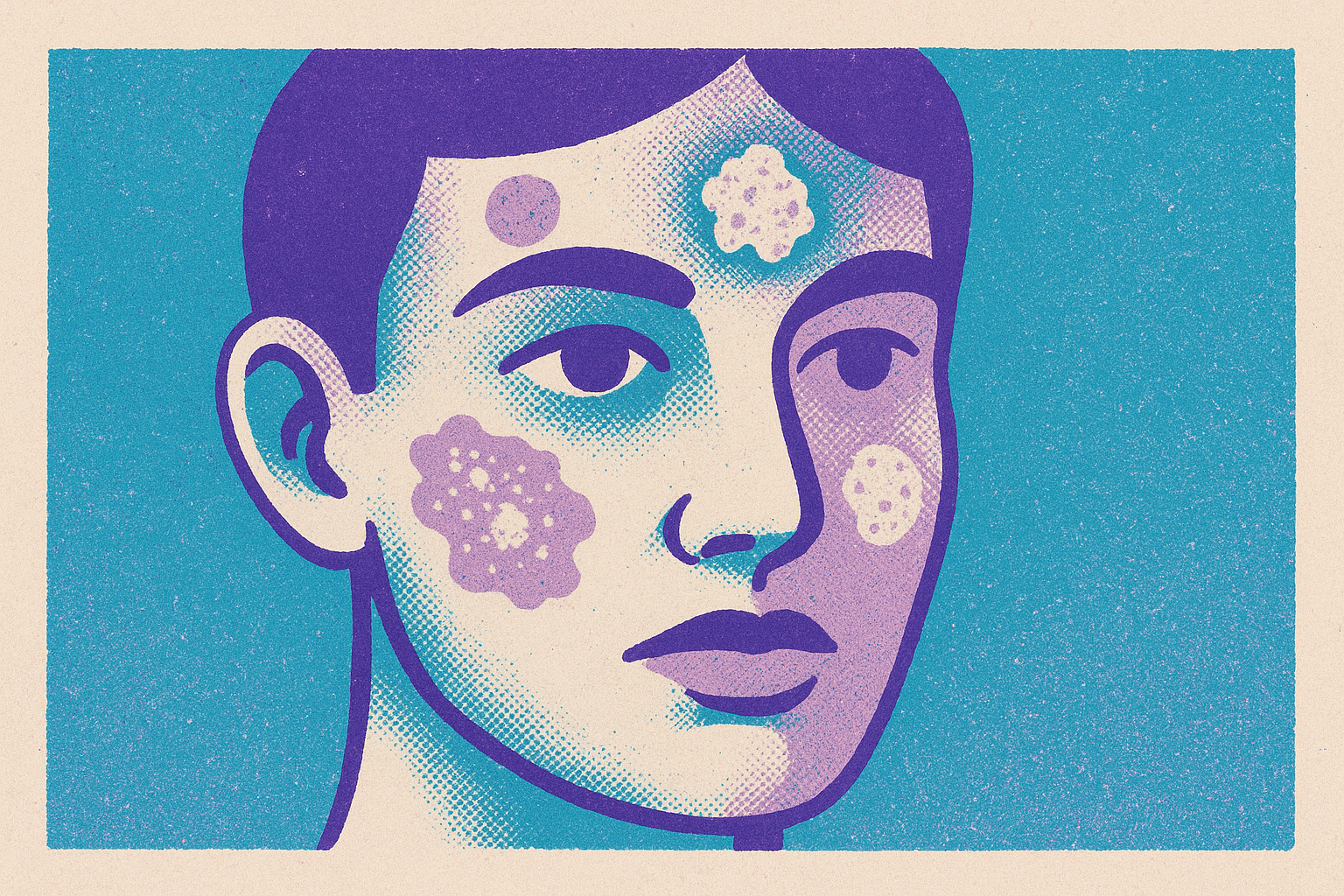
The face carries more years of sun exposure than almost any other part of the body, and that single fact explains why it shows up at the center of nearly every skin cancer conversation. Mayo Clinic lists the scalp, face, lips, and ears among the most common sites where skin cancer develops, and the reason comes down to decades of cumulative ultraviolet exposure hitting skin that rarely gets covered.
The epidermis holds three cell types, basal cells, squamous cells, and melanocytes, and each one gives rise to a different cancer with its own look, its own growth speed, and its own level of danger. That's the reason a single checklist can't cover all three. A bump that fits basal cell carcinoma looks nothing like a patch that fits squamous cell carcinoma, and neither looks like the kind of mole that signals melanoma.
Treating all three as one blurry category of "weird spot" is how early signs get waved off. Learning to tell them apart, one at a time, is the actual starting point for knowing when a spot on the face needs a dermatologist.
Basal cell carcinoma on the face
Basal cell carcinoma, or BCC, is the most common skin cancer overall, and the face is one of its favorite locations. MD Anderson describes it as a cancer that starts in the basal layer of the epidermis, which produces new skin cells. It tends to grow slowly and rarely spreads to other parts of the body. Left alone for long enough, though, it can invade and destroy the tissue and even the bone underneath it.
That combination is what makes BCC so easy to misjudge. A cancer with a low risk of spreading sounds, on the surface, like something that can be ignored for a while. But slow growth and low spread risk don't mean low stakes: the longer a BCC sits untreated on the face, the more local damage it can do to skin, cartilage, and bone in a part of the body with very little room to spare.
On the skin, MD Anderson's symptom guide points to three recognizable patterns: a pearly or waxy bump, a patch that looks flat, rough, or scar-like, and a sore that bleeds, never fully closes up, or keeps coming back after it seems to heal. That last pattern is the one most often rationalized away. A spot that scabs over, looks like it's resolved, and then reopens weeks later reads to most people as a stubborn pimple or a cut that won't quite finish healing, not as a tumor.
Skin tone changes what that bump actually looks like. Mayo Clinic notes that on white skin, BCC often appears as a skin-colored or pink bump, which matches most of the images used in public skin cancer education. On brown and Black skin, it's more often a brown or glossy black bump with a rolled border, a presentation that gets missed often, by patients checking their own skin and sometimes by clinicians too. A pigmented BCC can look enough like an ordinary mole that there's no obvious reason to flag it. Put together, slow growth, a healing-then-returning pattern, and a pigmented appearance that mimics a benign mole are three separate reasons the same cancer manages to sit on someone's face for months or years before anyone takes a second look.
Squamous cell carcinoma: the rougher, faster-moving cousin that behaves differently on darker skin
Squamous cell carcinoma, or SCC, starts in the squamous cells that make up most of the epidermis, MD Anderson explains, and it behaves differently from BCC in almost every respect that matters. It grows faster. It carries a higher chance of spreading. It's more likely to invade bone and muscle if it goes untreated. Where BCC rewards patience with slow, contained growth, SCC punishes delay.
The texture gives it away before the growth rate does. BCC tends to look smooth and pearly. SCC is rough, scaly, and prone to crusting or bleeding, sometimes developing a thickened, warty surface that doctors call hyperkeratosis. It can also ulcerate. That roughness is distinctive once someone knows to look for it, but it's also exactly the kind of thing that gets mistaken for a persistent patch of dry skin, a wart, or a scab that just won't quit. MD Anderson's symptom list includes a firm red nodule, a scaly or crusty lesion with irregular borders, and a lesion that's painful or itchy to the touch. Mayo Clinic flags the lower lip and the ears as especially common spots for SCC on the face, both places that take a steady beating from the sun over a lifetime.
The sun-exposure story gets complicated here. MD Anderson notes that SCC can also appear on skin that never sees the sun, and this pattern occurs especially often in people with darker skin tones. SCC makes up a disproportionately higher share of all skin cancers diagnosed in people with darker skin. That means the assumption that the face is the main site to watch doesn't hold equally for everyone. For some patients, the areas to check extend well beyond sun-exposed skin on the face and scalp.
Actinic keratosis, or AK, is a precursor that shows up as a red, scaly patch on sun-exposed skin, commonly on the face and scalp, caused by UV damage to the machinery inside cells that controls how they divide. Not every AK turns into SCC, but a share of them do, and catching one early gives a dermatologist a chance to treat it before it has the opportunity to progress. A rough, scaly patch that won't go away is worth a look specifically because it might be sitting at that earlier, more treatable stage.
Melanoma on the face and the ABCDE rule
Melanoma begins in melanocytes, the pigment-producing cells in the skin, and MD Anderson describes it as less common than BCC or SCC but considerably more dangerous, with the highest capacity of the three to spread to other parts of the body. Caught early, the five-year survival rate approaches 100%. That single number is the entire argument for learning to recognize it fast.
The ABCDE rule is the tool most dermatologists hand patients for exactly this reason, and it holds up well for the most common form of melanoma. Asymmetry means one half of a spot doesn't match the other. Border means the edges look ragged, notched, or blurred. Color means the spot isn't one uniform shade, it might mix brown, black, pink, red, white, or blue. Diameter flags anything larger than about 6 millimeters, roughly the size of a pencil eraser, though MD Anderson notes melanomas can be smaller than that. Evolving covers any change in size, shape, color, or behavior, including new itching or bleeding. Beyond the five letters, MD Anderson also lists a sore that won't heal, redness or swelling that spreads past a mole's border, color bleeding into the surrounding skin, tenderness or itching around a mole, and a texture change like scaling, oozing, or bleeding.
On the face specifically, a subtype called lentigo maligna shows up as a tan-brown to dark brown patch with an irregular border that grows slowly, most often on sun-exposed skin like the face and neck. Peak incidence falls between ages 65 and 80, and these lesions tend to run larger than other melanoma types, generally at least 6 millimeters wide. Left untreated, lentigo maligna can progress into a more invasive form called lentigo maligna melanoma. For an older adult with a slowly spreading pigmented patch on the face, this is the pattern to have checked.
The gap that matters most involves nodular melanoma. Nodular melanoma, the most aggressive form of the disease, breaks from the ABCDE pattern. Instead of an asymmetrical, multicolored, slowly spreading patch, it can show up as a uniform, raised bump that grows quickly, with none of the five warning signs the checklist is built around. Someone running through ABCDE and coming up empty could still be looking at the most dangerous version of this cancer. A lesion that's changing fast and doesn't match any familiar description deserves a dermatologist's attention even when it fails every box on the checklist. Mayo Clinic makes a related point: the first sign of melanoma is very often simply a mole that starts changing in size, shape, or color. Change itself, more than any single feature on a list, is the signal to act on.
Symptom differences across skin tones and late diagnosis
A pattern cuts across all three cancers: the visual signs described above are recognized less reliably on darker skin, by the people living in that skin and, at times, by the clinicians examining it. These cancers are diagnosed later and have worse survival outcomes as a result of that gap.
Mayo Clinic spells out the mechanism. In people with Black or brown skin, skin cancer tends to occur in places that don't get much sun: palms, genitals, under the nails. Watching the face closely is still worthwhile, but it isn't enough on its own for every patient. The same gap is visible at the level of individual cancers. BCC on darker skin tends to appear brown or glossy black with a rolled border, not the pink or skin-colored bump shown in most widely circulated patient education images. SCC occurs disproportionately more often in people with darker skin tones, and it can develop in places with chronic inflammation, old scars, or burns, sites nobody associates with sun damage and so nobody thinks to check.
These recognition gaps carry real weight. Skin cancer in patients with darker skin tones is frequently diagnosed at a later stage, and melanoma survival rates run lower in these populations. Skin cancer overall is about 30 times more common in non-Hispanic white people than in non-Hispanic Black people or people of Asian or Pacific Islander descent, a gap that comes from lower awareness, fewer routine skin exams, and cancers forming in spots that are harder to see or simply don't get checked. Lower overall incidence in a population does not translate to better outcomes when the disease does occur. It can mean the opposite, because awareness, screening habits, and visual pattern recognition all lag behind.
Knowing the brown-or-glossy-black presentation of BCC, or checking palms and nail beds as well as the face, is the difference between catching a cancer early and catching it late.
Physical sensations that accompany visible changes and the signs that have no pain at all
Waiting for a spot to hurt before worrying about it is one of the most common reasons people put off a dermatology visit, and it's also one of the easiest mistakes to understand once the biology is laid out. MD Anderson notes that skin cancer can cause itching, tenderness, pain, or a burning feeling, so sensation is sometimes part of the picture. But many skin cancers feel exactly like the normal skin around them, no pain, no itch, nothing to alert anyone through touch alone.
That absence of sensation is precisely why visual monitoring can't be optional. A pearly BCC bump might sit quietly for months without a hint of discomfort while slowly working its way deeper into tissue. A melanoma can itch or bleed as it advances, but plenty of early melanomas give no physical warning. SCC is more likely to announce itself through pain or itching alongside its rough, scaly texture, which gives it a slightly different profile from the other two. None of these sensations, present or absent, says anything definitive about how serious a spot is on its own.
Treat any new or changing spot on the face, painful or not, as information worth acting on rather than waiting for it to declare itself through discomfort. A sore that won't close, a patch that keeps changing shape, or a bump that's been sitting there for weeks earns a second look regardless of what it does or doesn't feel like. Pain can be a clue. Its absence is never a clearance.
Sources
- 9 Top Skin Cancer Symptoms & Signs
- Skin Cancer
- Skin cancer - Symptoms and causes - Mayo Clinic
- Skin Cancer: Symptoms & Causes
- Finding skin cancer in darker skin tones
- Skin Cancer in Skin of Color
- ABCDEFG of melanoma
- Risk Factors and Innovations in Risk Assessment for Melanoma, Basal Cell Carcinoma, and Squamous Cell Carcinoma - PMC