Squamous Cell Carcinoma Stages and Visual Progression
Early detection transforms squamous cell carcinoma from a simple office fix into a complex battle.
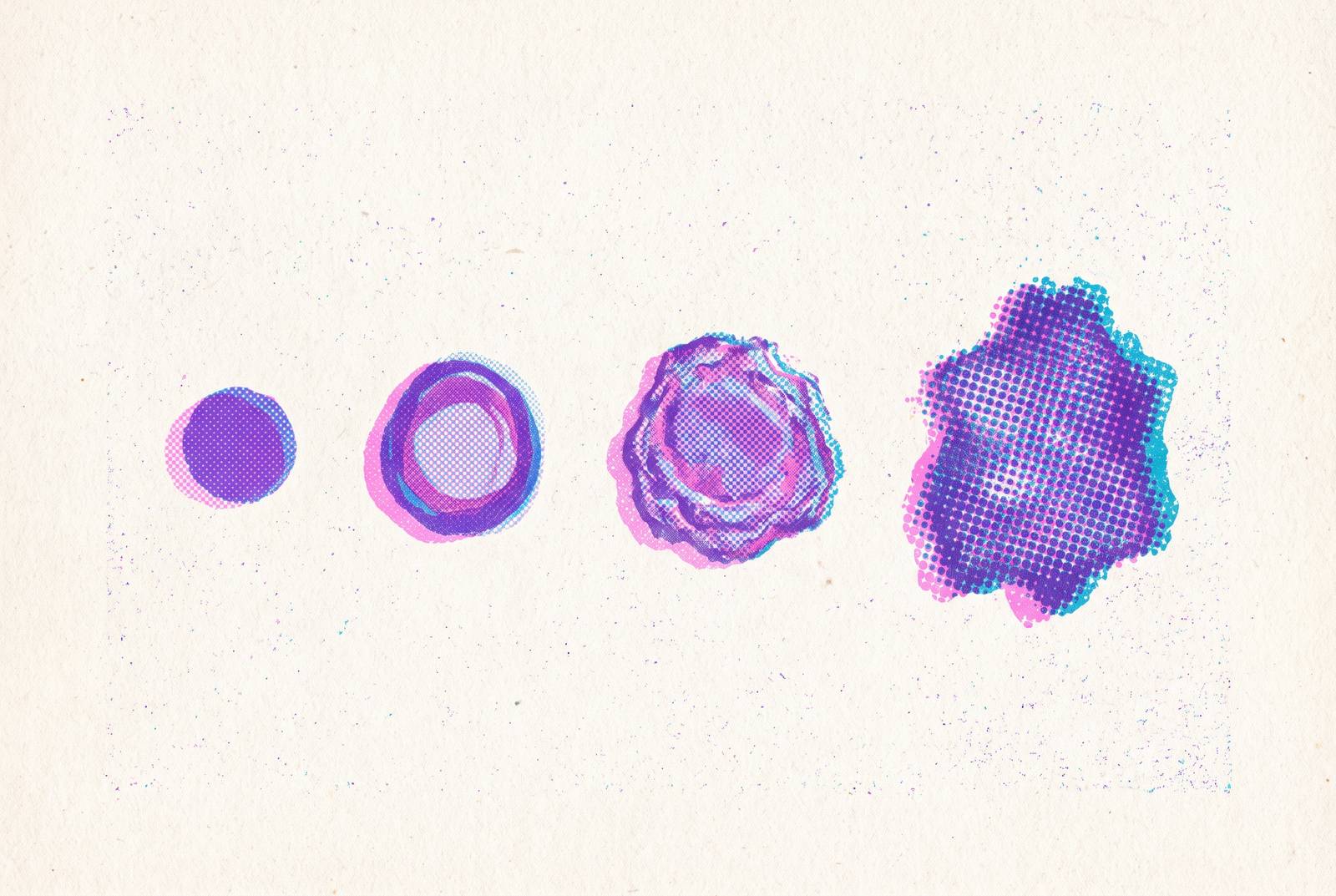
Squamous cell carcinoma starts small: a single flat cell in the top layer of skin, its DNA scrambled by years of UV exposure, dividing when it shouldn't. What happens after that first mutation, and what it looks like as it happens, is the difference between a five-minute office procedure and a fight for survival. This piece walks through each stage of that progression, from the earliest warning sign to distant spread, so the visual changes make sense as more than cosmetic detail.
Squamous cells are the flat, thin keratinocytes that make up the outermost layer of skin, the epidermis. They're built to shed and regenerate constantly, which is part of why they're so vulnerable: every UV exposure, every free radical event, chips away at their ability to repair DNA damage or self-destruct when something goes wrong. Malignant proliferation, cells growing and dividing when they should be dying off, is the event at the center of SCC. And it doesn't only happen on sun-exposed skin. SCC can develop in the mouth, the airways, the lungs, the digestive tract, even the urinary tract, though the visual signs and staging criteria shift depending on where it develops.
SCC is a spectrum, not a switch that flips from healthy to cancerous. It's a spectrum. Actinic keratosis progresses to intraepidermal carcinoma, which can progress to invasive SCC, and each step represents advancing stages of the same underlying disease. Actinic keratosis can regress on its own. Intraepidermal carcinoma often responds to immune-activating treatments. Invasive SCC typically doesn't reverse course once it's established. That asymmetry, the fact that earlier stages have an exit ramp and later ones don't, is the entire argument for catching this early.
As lesions move from precancerous to cancerous, the tissue's chemical signaling appears to shift from proinflammatory cytokine production toward chemokine production. That change in signaling helps explain why the biology, not just the size of a lesion, marks when the window for simple treatment starts closing. The rest of this piece works through what that looks like, stage by stage, on the skin.
How common SCC is and why incidence keeps rising
Squamous cell carcinoma is the second most common form of skin cancer, and it affects more than 1 million Americans every year. Lifetime risk is somewhere between 14% and 20% in the populations that have been studied, and that number is climbing. It's climbing.
How much? Estimates vary, but the trend line is consistent: incidence has risen by at least 50% over the past 30 years, and some analyses put the increase closer to 200% over that same period. Germany's numbers are even starker, with a reported fourfold increase over three decades. Among people of Caucasian descent, current incidence is around 31.4 cases per 100,000 annually.
Two forces are driving this, and neither is likely to reverse anytime soon. First, cumulative UV exposure: people are living longer, and skin damage compounds over a lifetime, so a population that's aging is a population accumulating more sun damage per person. Second, the aging population itself, since SCC risk climbs steeply with age. The American Academy of Dermatology estimates that roughly 90% of non-melanoma skin cancers, SCC included, trace back to UV exposure, a known cause acting on more people, for longer, than it used to. That's not a mystery disease. It's a known cause, acting on more people, for longer, than it used to.
So what does that mean practically? More people are going to encounter SCC in their lifetime, whether on their own skin or a family member's. Visual literacy, knowing what to look for and when it matters, becomes more valuable, not less, as the numbers climb.
The precursor lesion: what actinic keratosis looks like and why it matters
Roughly 82% of cutaneous SCCs start as actinic keratosis, a rough, scaly patch that appears on sun-exposed skin: face, scalp, hands, forearms. It's often the first visible sign that skin has taken more UV damage than it can fully repair.
Actinic keratosis has a fairly distinct feel, if not always a distinct look. The texture is the giveaway: rough, almost like sandpaper, even when the color barely stands out from surrounding skin. Sometimes it's pink or red, sometimes brown, sometimes it just blends in. That's part of the problem. It often feels more serious under a fingertip than it looks in a mirror.
Any single actinic keratosis lesion has a low chance of progressing to invasive cancer, around 0.24% per year. Any single actinic keratosis lesion has a low chance of progressing to invasive cancer, around 0.24% per year. That sounds almost negligible. But most people with actinic keratosis don't have just one lesion, they have several, and risk compounds at the patient level. Looked at over ten years, with multiple lesions present, progression risk climbs to somewhere between 6.1% and 10.2%. For the more severe, treatment-resistant cases (what's classified as Olsen grade III), progression risk can run as high as 33.5%.
The practical problem is that actinic keratosis looks a lot like ordinary dry skin, or garden-variety sun damage, or just a rough patch that won't quite go away. That visual ambiguity is exactly what delays people from getting it checked. And it's the same ambiguity that carries forward into the next stage, where the stakes go up but the appearance barely changes.
Stage 0 (carcinoma in situ): confined to the surface, but not safely ignorable
Stage 0 SCC, sometimes called carcinoma in situ or Bowen's disease, means abnormal cells are present but haven't broken through the epidermis yet. No invasion into deeper tissue. Structurally, this is as early as SCC gets.
Visually, it tends to show up as a flat, rough, scaly patch, pink, red, or close to skin tone, with a texture that can flake or crust when touched. In its earliest form, it can spread into a fairly large reddish patch, sometimes over 2.5 cm across, with borders that are well-defined but often irregular in shape. Most commonly it appears on sun-exposed skin: face, ears, neck, the backs of hands, forearms. But it isn't limited to those zones. It can show up on covered skin too, and in the genital region, where it's often linked to a specific virus rather than sun exposure.
What makes Stage 0 tricky isn't rarity, it's mistaken identity. It gets confused for eczema, psoriasis, ordinary dry skin, or actinic keratosis constantly. That overlap isn't a minor inconvenience, it's the main clinical danger at this stage. A patient sees a rough patch, applies moisturizer, watches it seem to calm down, and moves on. Then it comes back, maybe slightly different in shape or color. That return, that persistence, is the actual signal. Not pain, not rapid growth, just a patch that won't fully go away and quietly changes shape over weeks or months.
Location changes the picture too. In the mouth, Stage 0 SCC can look like white or red patches. In the eye area, it might just cause itching or burning, nothing visibly dramatic. This is, without much argument, the most treatable stage SCC ever passes through. Nailing down what it looks like determines how early it can be caught and treated, since this is the most treatable stage SCC ever passes through.
Stage 1: the lesion becomes invasive but is still small and highly curable
Stage 1 marks the shift from "confined to the surface" to genuinely invasive: cancerous cells are now present within the skin itself, though the tumor stays small and hasn't reached the lymph nodes.
Physically, that tends to look like a small, firm, raised bump or plaque, generally under 2 cm across. The surface might be rough, crusted, or scaly. Color runs pink to red, sometimes a shade darker than the skin around it. Some lesions develop a central depression or minor ulceration, and might bleed if bumped or scratched. Common sites include the face, ears, lower lip, scalp, and the backs of the hands, the same sun-battered zones actinic keratosis tends to favor.
It's easy to write this off as a stubborn pimple, a rough patch of keratosis, or a small wound that's just slow to heal. The raised texture is new, but nothing about it screams alarm on its own, which is exactly the danger.
In TNM staging terms, this is T1: a small, localized tumor with no node involvement and no distant spread. Localized, in other words, in the most literal sense.
Surgical treatment at Stage 1 and Stage 2 succeeds more than 95% of the time, which is about as good as outcomes get in oncology. Surgical treatment at Stage 1 and Stage 2 succeeds more than 95% of the time, about as good as outcomes get in oncology. That's about as good as outcomes get in oncology. But "small" doesn't mean "safe to wait on." Acting at Stage 1 is precisely what keeps treatment this simple.
Stage 2: the lesion grows larger and deeper, raising the stakes for treatment
Stage 2 SCC has grown into deeper layers of skin, though for cutaneous disease, it still hasn't reached the lymph nodes. Size is usually the visible tell: lesions cross the 2 cm mark, turning into larger raised growths, wart-like bumps, or open sores that refuse to heal. The surface often crusts, ulcerates, or bleeds on its own, and surrounding skin can look inflamed or irritated.
But size isn't the only path here. Certain high-risk features can bump a smaller lesion into Stage 2 territory regardless of its measurements: perineural invasion (cancer tracking along a nerve), significant tumor depth, location near the eye, ear, or lip, or a patient who's immunosuppressed. Any one of those changes the calculus even on a lesion that looks unremarkable.
Symptoms tend to catch up with appearance at this stage. There's more discomfort, more visible irritation, more reason for a patient to actually notice something's wrong. Location adds its own risk multiplier: facial SCC near the eye, ear, or lip gets classified as high-risk no matter its size, because those areas have thin tissue planes and not much room for a wide surgical margin.
In TNM terms, this is T2: a tumor larger than 2 cm, or one carrying multiple high-risk features regardless of size. Treatment options widen here too. Mohs micrographic surgery remains the preferred approach for high-risk locations, since it allows tissue-by-tissue margin checking during the procedure itself. Standard excision, radiation, cryotherapy, and electrodesiccation stay on the table for lower-risk presentations. The window for treatment hasn't closed at Stage 2, but the procedure involved is bigger, the margins tighter, the stakes higher. That's the argument for acting sooner rather than later, made concrete.
Stage 3: regional spread changes both what the patient sees and what treatment can do
Stage 3 is where SCC crosses a real threshold: it has either reached nearby lymph nodes or grown further into deeper structures like bone or muscle. This is regional disease, not yet distant, but no longer confined to the original site.
Physically, the primary lesion tends to be sizable, often substantial in size for cutaneous SCC, though a smaller lesion can still qualify for Stage 3 if regional lymph node involvement is present. The lesion may be ulcerated, or fixed in place, no longer moving freely over the tissue beneath it, a sign it's anchored into deeper structures. Nearby lymph nodes might become visibly or palpably swollen, which is a genuinely new sign, not present at any earlier stage. Skin around the lesion can show tethering or a loss of its normal contour, sometimes with satellite lesions nearby. The tumor may also extend into fat tissue below the skin.
For oral SCC specifically, Stage 3 can start interfering with swallowing, particularly if the tumor sits near the base of the tongue, the throat, or the esophagus. Patients sometimes describe feeling a lump in the throat that wasn't there before. Symptoms, in other words, start carrying functional weight, not just cosmetic concern.
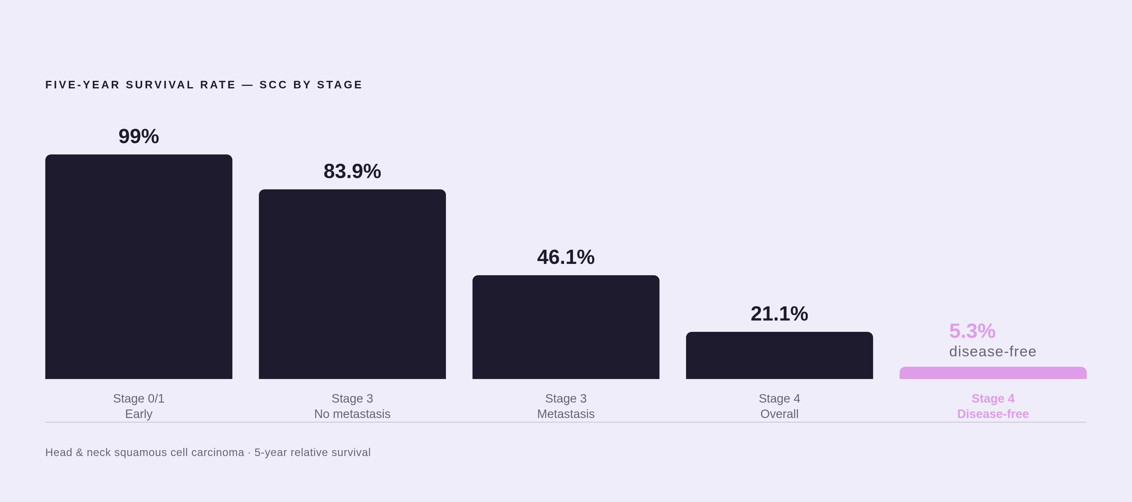
Treatment shifts accordingly. Local removal is typically combined with adjuvant radiation therapy, and for higher-risk cases, systemic therapy may run concurrently with radiation to cut recurrence risk. What does that combination buy a patient? Prognosis data gives a blunt answer: five-year survival is 83.9% for patients without metastases, but drops to 46.1% for those with metastases. That gap, nearly forty percentage points, is the clearest argument in this entire progression for catching disease before it reaches this stage.
Certain groups face a higher likelihood of reaching Stage 3 in the first place, particularly those who are immunosuppressed. For these groups, catching a lesion depends on more frequent monitoring, not just general awareness.
Stage 4: what distant metastasis looks like and what it means for prognosis
Stage 4 means the cancer has reached one or more distant organs, commonly the lungs, bone, brain, or liver. At this point, the disease has left its original neighborhood entirely.
On the skin, the primary lesion is typically large, deeply ulcerated, and can invade underlying bone or cartilage, producing a destructive, crater-like wound that simply won't heal. But the more dangerous part of Stage 4 isn't visible on skin exam at all. Distant spread instead produces systemic symptoms: fatigue, unexplained weight loss, night sweats, wheezing or persistent coughing, trouble swallowing, even changes in personality or cognitive function if the brain is involved. The disease, by this point, is no longer something a patient can simply look at and assess.
Five-year survival is 99% when SCC is caught early. For advanced disease, that number drops well below 50%. For metastatic head and neck SCC specifically, five-year overall survival is 21.1%, and disease-free survival is just 5.3%. That's the widest gap in this entire progression, and it's the clearest evidence that timing, more than almost any other factor, determines outcome.
Metastasis itself is uncommon, affecting roughly 2% of all cutaneous SCCs. But when it happens, it tends to happen early: a JAAD registry study tracking 11,137 patients found a median time to metastasis of 1.5 years from diagnosis, with about three-quarters of cases occurring within the first four years. That's not a slow-moving risk sitting in the background for decades. It's a relatively narrow window, which makes the case for early-stage vigilance even stronger. None of these numbers are meant as a verdict. They're the argument for showing up to a dermatologist at Stage 0 or Stage 1, when the odds are still overwhelmingly favorable.
Why SCC on the face, scalp, and other high-risk sites behaves differently across all stages
Stage alone doesn't tell the whole story. Location acts as its own independent risk factor, and it shapes both appearance and danger at every stage above.
Take the face and scalp: SCC here carries a higher recurrence risk than SCC on the trunk or limbs, and facial lesions near the eye, nose, or ear get flagged as high-risk regardless of size, purely because there's so little tissue to work with surgically. The lower lip presents its own pattern, a persistent scaly patch or erosion that won't heal, sometimes with an increasingly irregular lip border. It's frequently written off as sun exposure or ordinary chapping, which delays evaluation. The ear, particularly the outer rim, tends to develop a firm, flesh-colored or reddish plaque that's often painless early on. That absence of pain is deceptive: thin tissue planes near the ear mean even a small tumor carries elevated risk. The nose and nasal rim show raised, crusty nodules prone to bleeding with light contact, while the temple and forehead, both chronically sun-damaged zones, tend to develop scaly, indurated plaques.
Leg and scalp presentations look visually distinct from facial SCC, which reinforces a broader point: SCC doesn't have one universal appearance. Scalp SCC in particular is easy to miss since hair conceals it, and it's frequently mistaken for seborrheic dermatitis or ordinary dandruff.
Any lesion near the eye, ear, or nose deserves earlier surgical evaluation, not a wait-and-see approach. Proximity to those structures limits what can be safely removed later, so delay costs more in these zones than it does elsewhere on the body. And genital SCC deserves its own mention here too, since it's often HPV-related rather than sun-driven, and appears in covered skin rather than sun-exposed areas. That's a useful reminder that UV exposure is the dominant driver of SCC, but not the only one, and self-monitoring needs to cover more ground than just the obviously sun-exposed parts of the body.
Risk factors that accelerate progression and who faces the highest burden
Certain groups carry a disproportionate share of SCC risk. Fair-skinned individuals, particularly those of Caucasian descent, face higher baseline incidence, largely tied to lower natural UV protection in the skin. Cumulative sun exposure over decades drives risk more than any single sunburn, which is part of why age is such a consistent risk driver: more years alive generally means more accumulated UV damage.
Immunosuppression stands out as one of the sharpest accelerants across every stage discussed here. Patients on immunosuppressive medication, whether for organ transplants or autoimmune conditions, face not just higher SCC incidence but faster progression once a lesion forms. That's consistent with the pattern seen earlier: immunosuppressed patients appear repeatedly as a higher-risk group, from actinic keratosis progression through Stage 3 regional spread.
Men also carry a higher burden than women, and that gap widens with age, an important detail for that group in particular to sit with rather than gloss over. Occupational and lifestyle sun exposure compounds this further: outdoor workers, and anyone with a long history of sunburns rather than protective habits, accumulate risk at a faster clip than someone whose exposure has been lighter or better protected.
None of these risk factors are exotic. They're well-documented, consistent, and, in most cases, at least partially modifiable through sun protection and regular skin checks. What separates them from something you can meaningfully act on is simply attention, catching a lesion at Stage 0 rather than Stage 3, and knowing which patches of skin deserve a second look rather than a shrug.
Sources
- Squamous Cell Carcinoma Stages Pictures: Full Visual Guide
- Global status of research on cutaneous squamous cell carcinoma and its programmed cell death: Bibliometric and visual analysis from 2012 to middle 2022 - PMC
- Cytokine/chemokine profiles in squamous cell carcinoma correlate with precancerous and cancerous disease stage - PMC
- pmc.ncbi.nlm.nih.gov
- theminorsurgerycenter.com