Why Consistent Skin Photo Documentation Matters
Photos turn guesswork into objective evidence your dermatologist can act on.
Skin conditions don't announce themselves overnight. Eczema spreads over weeks, a mole darkens over a year, acne cycles with the seasons, and none of that shows up on a timeline memory can hold onto. I've sat with patients who swear a spot "just appeared last week" when the photo on their own phone puts it there two months earlier. Memory rounds off. Cameras don't.
Here's the actual problem with relying on memory: people don't compare "now" to an accurate "then." They compare it to whichever moment felt most vivid, usually the worst flare or the one time their skin looked great, and that comparison is almost always skewed. I've had patients sit down and genuinely not know when something started, whether it's better than last month, or what happened right before it flared. Those aren't small gaps. The trajectory of a skin change often points toward the diagnosis as much as the change itself does. A photo is what fills that gap. Call it a memory prosthetic if you want a phrase for it; the reason it works is simple enough, a camera doesn't round things off the way a brain does.
What dermatologists actually photograph and why the practice is so widespread
Clinical photography isn't some niche habit tucked into one corner of dermatology. It's baked into how the whole specialty runs. A study out of Korea that combed through 111,426 photos in a dermatologic image archive found that marking biopsy sites was the single most common reason a photo got taken. But that's just one entry on a longer list.
We photograph to track disease progression. We photograph to check whether a treatment is doing anything. We photograph so a colleague can weigh in without the patient booking a second appointment. We photograph pigmented lesions that might be turning into something worse, and we photograph for teledermatology visits where, frankly, the photo often is the visit.
A verbal description of a rash, no matter how careful, can't sit next to last month's version of itself the way an image can. And the conditions we track this way the most, acne, psoriasis, atopic dermatitis, rosacea, happen to be the ones that fluctuate the hardest. Acne alone touches something like 85% of people between 12 and 24. These aren't conditions where one glance tells the whole story. They move, and a photo is how you catch the motion instead of guessing at it after the fact.
Dermatologists have leaned on this for decades. What's changed is that patients now carry the same tool around in their back pocket.
How regularity turns an individual photo into a usable timeline
One photo tells you what something looked like on one particular Tuesday. That's it. A series of photos, taken the same way at regular intervals, tells you what something is actually doing, which is a completely different category of information.
Regularity is the thing that makes the comparison mean anything. Same lighting, same angle, same distance, these aren't fussy details for the sake of tidiness. They're what separates a real change from a lighting artifact. Move the camera six inches closer and a patch looks bigger. Shoot under a warmer bulb and it looks darker. That's not biology; that's a setup problem dressed up as a symptom.
Get the setup consistent, though, and a timeline starts showing you things no single photo ever could. Is this a slow creep or a sudden jump? Does the flare follow a monthly rhythm, or does it track the seasons? Is the overall arrow pointing up, down, or sideways? You can even line up a new product, or a brutal week at work, against a flare that shows up two weeks later and start asking whether the two are connected.
There's a study that backs this up in a way I find genuinely convincing. In a year-long psoriasis self-monitoring project, 50 patients tracked severity, quality of life, and stress through a smartphone app, with a dermatologist checking in on the data every two months. What patients reported through the app lined up closely with what the physician found on exam. Regularity is what turned a stack of personal photos into something a clinician could actually work with, not just something a patient kept for their own peace of mind.
What a good photo actually captures, and the common mistakes that quietly ruin it
A useful clinical photo does two jobs at once. It gets close enough to show texture and color, and it holds enough surrounding skin in frame that anyone looking at it six months later can tell where on the body they're looking.
A few things make that possible, and none of them are complicated. Lighting has to stay consistent: natural daylight or a neutral bulb, never a flash (it washes out color), never warm indoor light (it distorts it). Distance has to stay fixed, close enough for detail, wide enough for context, and it's worth taking a wide reference shot first before zooming into the close-up. Angle needs an anchor, a floor tile, a doorframe, a specific spot to stand in front of a mirror, something that keeps the frame from drifting week to week. And filters, auto-smoothing, any of the "beauty" modes most phone cameras default to now: skip all of it. Those modes quietly adjust color and texture in ways that undercut the entire point of the exercise.
The mistakes that wreck a timeline are almost all avoidable, which is what makes them frustrating. Flash reflection is one. Shifting distance between sessions is another, since it can make a lesion that hasn't moved an inch look like it's growing. Only pulling out the camera when the skin looks bad introduces a selection bias that skews the whole baseline. And skipping the date on a photo means nobody, not you, not your dermatologist, can reconstruct the timeline later.
A 2024 study that reviewed 100 questionnaires on patient-taken smartphone photos of atopic dermatitis zeroed in on image quality and what the researchers called patient "digital readiness." Teaching patients how to photograph their own skin correctly is turning into a real clinical priority, not some afterthought tacked onto a visit. Even brief instruction on technique has been shown to move diagnostic outcomes in teledermatology. Method counts here. It's not just about meaning well.
How a photo timeline changes what a clinician can do between visits
Most skin conditions get managed across a string of visits spaced weeks or months apart. Everything that happens in between is invisible to the clinician unless the patient shows up with proof. That's the gap a photo timeline closes.
With a decent set of images, a clinician can tell whether a treatment is working before the next scheduled appointment, instead of guessing during a rushed five-minute check-in. They can catch a worsening trend early enough to actually do something about it. They can tell a passing flare apart from real progression. And they can take a reported trigger, a new lotion, a stretch of bad sleep, and check it against what the skin actually did afterward, rather than taking the patient's word for it alone.
This is the entire premise behind asynchronous, or store-and-forward, teledermatology: a clinician reviews photos and history a patient submits, then responds, usually within 24 to 72 hours, no live visit required. Diagnostic agreement between teledermatology and in-person exams sits at a mean kappa of 0.57, moderate to good agreement, and image quality moves that number around more than you'd think. Better photos, better agreement.
Consider how long people are waiting for care right now. Dermatology wait times are well documented as a persistent problem, and patients routinely report that their condition worsens during the gap before they're seen. A photo timeline doesn't shrink the waitlist. It won't get you seen faster. But it's one of the only things a patient can actually control that makes the appointment, whenever it finally lands, worth more once it happens. Photos don't replace the visit. They make the visit count for something.
Where AI-assisted review fits into this picture, and where it still needs a clinician
AI tools for reading skin images are real and improving fast. How well they work, though, depends entirely on what you're asking them to do.
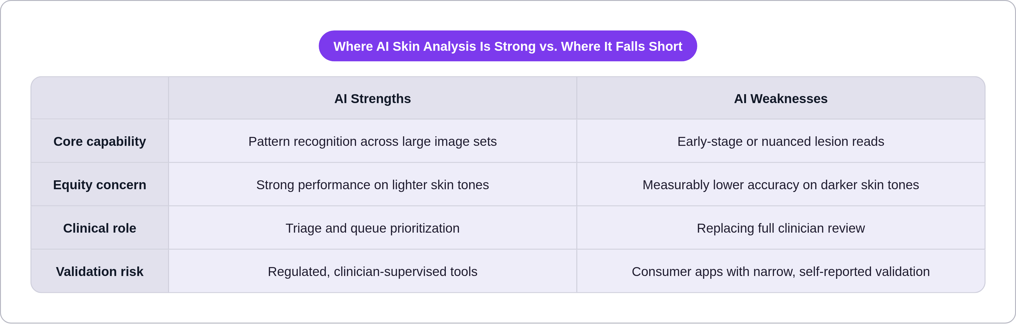
On the strong side, pattern recognition across huge sets of images is something these models are genuinely good at. CNNs trained on datasets like ISIC land somewhere in the 82% to 95% range for classification accuracy on common lesion types. Some commercial platforms report 91% to 94% in their own validation work. AI is also useful for triage: flagging which photos need a clinician's eyes today versus which can sit in a queue for a week.
Where it gets shaky is worth sitting with for a second. Early-stage nodular melanoma is one soft spot; one analysis found accuracy dropping to 68% at exactly the stage where catching it early matters most. Skin tone equity is another, and this one bothers me more than the melanoma number does. A 2025 meta-analysis found a pooled AUROC of 0.89 for lighter skin tones (Fitzpatrick I through III) against 0.82 for darker skin tones (Fitzpatrick IV through VI). That gap lands squarely on populations already underserved in dermatology. It's not a rounding error, it's a pattern. And consumer apps trained on small, narrow image sets, validated only by the company that built them, shouldn't get treated as clinical-grade just because the marketing uses clinical language.
The WHO's 2025 digital health assessment flagged three recurring problems in skin AI tools: a disconnect between the image analysis and the actual clinical reporting workflow, thin patient education built into the product, and confidence scores that spit out a flat yes-or-no with none of the nuance a real read requires.
So where does that leave the photo you took this morning? AI can help flag a change worth a second look. But reading a full timeline, deciding what a change actually means, deciding whether a treatment plan needs to shift, still takes a licensed clinician who can see the whole picture and the whole patient in front of them. Clinician-supervised AI, where the model backs up the person doing the reviewing instead of replacing them, is the direction both the evidence and the regulatory guidance are pointing.
Building the habit: what a realistic photo documentation routine looks like
How often you should photograph depends on what you're tracking. An active flare or a new lesion is worth a weekly photo, sometimes more. A stable, managed chronic condition is fine on a monthly rhythm. Mole or pigmented lesion monitoring usually runs on a three-to-six-month cycle, or whatever your clinician recommends. Starting a new treatment calls for a baseline photo before the first dose, then steady check-ins after.
Building the habit comes down to anchors, mostly. Tie it to something already happening on a schedule, the first of the month, a prescription refill, rather than trying to remember it cold. Stand in the same spot in the same room and you've already removed one variable before it becomes a problem. A folder or album labeled by condition keeps the photos from vanishing into a camera roll full of everything else. And a short note attached to each one, how the skin feels, anything unusual that week, what you're currently using on it, gives a picture context it can't carry on its own.
What you bring to the appointment matters too. Not everything you've shot, a curated set: the baseline, the worst point, where things stand now. Dates need to travel with the images; a clinician needs to know when a photo was taken almost as much as what's in it. For an asynchronous telehealth visit specifically, a clear, dated series paired with a short written history can cover most of what an in-person exam would otherwise gather, which is the model behind services like Nolla, an AI-guided online skin clinic where licensed US clinicians review submitted symptoms and issue prescriptions for acne, rosacea, and related conditions in about ten minutes.
Reviews of smartphone apps built for tracking acne, psoriasis, rosacea, and mole monitoring keep landing on the same thing patients actually care about: ease of use, simple tracking, nothing fancy. The technology was never the obstacle. The habit is. Stand in the same spot, use the same light, write down the date, and do it again next month. That's most of it.