How AI Analyzes Skin Images for Condition Detection
AI skin analysis tools rely on training data quality, and most struggle with darker skin tones.
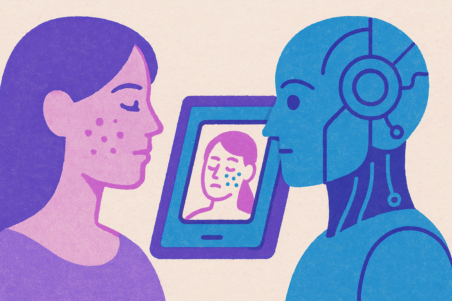
A patient who snaps a photo of a mole and runs it through an app is making a medical decision, whether they think of it that way or not. That difficulty is part of why people now open an AI tool before they call a doctor, since consumer apps and general-purpose chatbots are sitting right there on a phone, and a September 2026 literature review in JID Innovations points out that patients often reach for these tools before they ever seek formal care. Knowing what the model is actually doing, and what it can't do, puts a patient in a much better spot to judge when an AI result is good enough and when it's time to get a clinician to look.
How a convolutional neural network reads a skin image
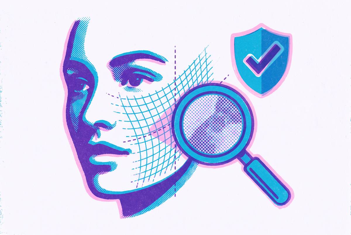
The technology behind almost every skin-analysis tool is a convolutional neural network, or CNN. Think of it as a machine that builds understanding in stages, the same way someone might describe a painting first by its colors, then by its shapes, then by what it actually depicts. A CNN starts with the simplest visual signals, like edges and color changes, then combines those into more complex features, like a jagged border or an asymmetric patch of pigment, and finally maps those combined features onto a diagnosis probability, all through layers it learned from labeled examples rather than hard-coded rules.
It helps to walk through what each stage is actually doing. The first layers of the network scan for pixel-level patterns, a line here, a shift in color there, across the whole image without caring where in the frame the pattern shows up. The next layers start stitching those simple signals together into something more specific: a rough border, a patch of uneven texture, a surface that looks scaly or crusted. By the final layers, the network compares the compound pattern it has built against thousands of similar patterns it saw during training, assigning a probability to each possible condition rather than thinking in terms of "mole" or "rash" the way a dermatologist does.
That process has measurable strengths. A 2025 review cited in Skinive's 2026 accuracy report found CNNs reaching 91% sensitivity when the task is distinguishing melanoma from benign lesions, which is the highest performance reported for that specific, high-stakes task. But notice what that number actually describes: it's a measure of how the model performs on a defined task under study conditions, not a guarantee about any single photo a patient takes at home. That distinction will matter more as this piece goes on.
What the training data determines
Everything a CNN can do traces back to what it was shown during training. A model's usefulness in the clinic is tied directly to the quality and range of its training images, and a model can be trained on millions of photos and still have no idea what to do with a presentation it never saw. That's the pivot point for understanding every claim that comes later in this piece: once you know how the model reads an image, the next question is what it was ever given to read.
Training works by feeding the network large volumes of labeled images, where a clinician or dataset curator has already confirmed the diagnosis attached to each photo. During training, the model just needs to find which pixel patterns reliably predict which labels. Skinive's accuracy report says the algorithm was trained on millions of skin images, and a clinician-validated subset of 250,000 was chosen specifically for training. That's a meaningful design choice: it shows the field moving toward verified, dermatologist-confirmed ground truth rather than just piling up raw image counts.
But volume and verification don't automatically produce range. A dataset built from real-world smartphone photos, taken under messy lighting at odd angles, teaches a model to handle the conditions patients actually photograph in. A dataset built entirely from polished clinical photography teaches something narrower. The JID Innovations review names restricted dataset sizes, limited diagnostic diversity, and selection bias as recurring limitations across AI dermatology studies, alongside inconsistent evaluation methods from one study to the next. And most of the AI models published between 2017 and 2025 were built to classify a specific, narrow set of conditions, often in binary form, like melanoma versus benign nevi. A model trained that narrowly is a genuinely different tool than one trained across hundreds of conditions, even if both get called "skin AI."
The deployment pipeline runs through three steps in sequence: data capture, model inference, and clinical review. Whatever limits exist in that first step set the ceiling for everything that follows. A model can't infer its way past a gap in what it was trained on, and a clinician reviewing its output is only as well-informed as the output allows.
What controlled accuracy figures leave out
Accuracy numbers from validation studies are real measurements, but they measure something narrower than you'd assume as a patient. They describe performance on a curated dataset, under conditions set up by researchers, not performance on the blurry photo someone takes in a bathroom with bad lighting of a condition the model has rarely seen.
That said, a fixed validation set is still a legitimate, reproducible way to track progress. The Skinive algorithm was tested against the same fixed validation set across five years and showed steady, measurable improvement by 2026. DermEngine and SkinVision, among other commercial platforms, report accuracy in the 91 to 94% range in their own commercial validation studies, consistent, trackable figures. What they don't settle is how the same model performs once it leaves the validation set and meets the full variety of real patients, real cameras, and real lighting. The JID Innovations review flags exactly this gap: strong controlled performance may not reflect real-world ability, and inconsistent evaluation is a problem across the literature.
The clearest way to see what a single accuracy number hides is to look at sensitivity and specificity side by side. Sensitivity measures how well a tool catches real cases; specificity measures how well it avoids false alarms. The FDA-authorized DermaSensor device achieves very high sensitivity across all skin cancer types, but very low specificity. It's a deliberate design decision for a tool meant to screen broadly in primary care, where missing a real cancer is a far worse outcome than flagging something that turns out to be benign. A single blended "accuracy" figure would flatten that trade-off into a number that tells a patient almost nothing useful.
One might ask where current systems are weakest, given all this. Early-stage nodular melanoma is the presentation current AI systems detect least reliably, which is also the presentation where catching it early matters most. The gap between reported accuracy and real-world reliability is narrowest for easy cases and widest for the cases that carry the highest stakes.
How skin tone bias emerges from training data
The most well-documented failure in AI skin analysis isn't a coding error. A structural imbalance is baked into the training data, so it produces measurably worse performance on darker skin tones, and that hits patients who already face the biggest gaps in dermatology access.
The mechanism traces straight back to the dataset imbalance raised earlier. The ISIC dataset, the field's primary public training resource, skews heavily toward Fitzpatrick types I through III, while types V and VI combined make up only a small share of all images in it. A model trained mostly on light skin learns patterns that are specific to light skin, and that gap turns into a measurable clinical consequence: one documented case found a dramatic drop in diagnostic accuracy for Black skin conditions compared to Caucasian images, using an AI dermatology application trained on Caucasian datasets. If a model trained on a standard dataset is tested against a curated darker-skin dataset instead, melanoma detection sensitivity drops severely, so performance degrades exactly where the consequences of a missed diagnosis are most serious.
When this happens, explainable AI techniques can show what's going on inside the model. When analyzing darker skin, models trained on biased data tend to focus on the skin surrounding a lesion rather than the lesion itself. The model isn't reading the same clinical signal a dermatologist would be reading. This isn't limited to purpose-built dermatology tools, either. Evaluations of ChatGPT-4o across the Fitzpatrick scale found significantly lower sensitivity, specificity, and accuracy for melanoma in darker tones, Fitzpatrick types III through VI, compared to lighter tones, types I and II.
The geography of this problem deserves attention on its own. A September 2025 systematic review in the International Journal of Emergency Medicine focused on low- and middle-income countries, precisely where dermatologist shortages run deepest and where skin presentations on darker skin are most common. A July 2025 editorial in International Health, covering AI and neglected tropical diseases, adds a sharper picture: many African countries have fewer than one dermatologist per million people, and those few are concentrated in cities, while the heaviest burden of skin disease is in rural communities. AI tools trained mostly on lighter skin are rolling out into exactly that gap. The places where dermatology access is thinnest are also the places where current AI performs worst, and that overlap isn't a coincidence; it's a direct result of who was and wasn't represented in the training images.
The clinical takeaway follows directly from all of this. A negative AI result for a patient with Fitzpatrick type IV, V, or VI skin should never be treated as the final word over a clinician's own suspicion, because the model's confidence was never calibrated for that patient in the first place.
Regulated devices, commercial platforms, and consumer AI in the landscape
Patients encounter a wide range of tools under the single label "AI skin analysis," and those tools sit in very different places on a spectrum running from FDA-authorized medical devices to general-purpose chatbots. Where a tool sits on that spectrum determines what claims about it can actually be trusted and how much weight its output should carry.
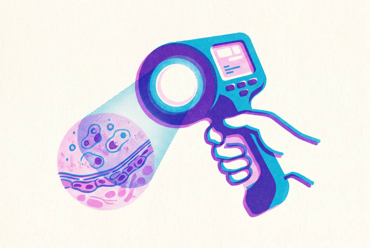
At the top of that spectrum sits DermaSensor, which received FDA authorization on January 17, 2024, making it the first AI-enabled medical device cleared in the United States for skin cancer detection by non-specialists in primary care. It had already been authorized in the EU, Australia, and New Zealand. DermaSensor uses spectroscopy rather than image analysis, sending light pulses into a lesion and reading the result.
Commercial platforms occupy the next tier. DermEngine and SkinVision use ensemble classification methods and give users basic reporting features, and they draw their accuracy figures from their own commercial validation studies. These tools are useful and well-documented, but they operate under a different regulatory standard than an FDA-cleared device.
A third category is in active clinical trial territory. Legit.Health, built by AI Labs Group S.L., ran a completed clinical trial from June through September 2024, registered with ClinicalTrials.gov, studying whether its AI medical device could help healthcare professionals diagnose rare and complex skin conditions more accurately, including generalized pustular psoriasis. It's a specific, narrow research question, tested through the formal clinical trial process, not a finished consumer product claim.
Then there's consumer-facing general-purpose AI. ChatGPT-4o is multimodal and available to anyone before they ever see a clinician, but its diagnostic accuracy for neoplastic, inflammatory, and vascular skin diseases started as low as 56% across several studies and didn't climb much past that, well under the validated accuracy of tools built specifically for dermatology.
And below all of that sits a large, mostly unregulated layer of smartphone apps. A 2024 analysis in JAMA Dermatology identified 41 dermatology AI applications available for download, none of them FDA-cleared, and only 2 of the 41 including any disclaimer about their regulatory status. A patient scrolling an app store has no visual way to tell those apart from a cleared medical device.
The regulatory environment itself is shifting quickly. The FDA has been authorizing AI-enabled medical devices at a growing pace, and a small share of 2025 clearances included a formal mechanism that lets a model's algorithm evolve after clearance within boundaries the FDA pre-approves. But a 2025 cross-sectional study in JAMA Network Open, examining FDA-authorized AI-enabled devices broadly, found limited public detail on clinical generalizability, validation scope, and the demographic diversity of the populations these devices were tested on. Regulatory clearance is a meaningful signal, but it isn't a complete window into how a device was tested or on whom.
What AI assistance does to clinician judgment
All of this eventually lands in an exam room, where a clinician is deciding how much weight to give an AI output. AI assistance raises diagnostic accuracy on average, but it carries a specific risk alongside that benefit: a clinician handed inaccurate AI output can end up performing worse than if no AI had been involved at all, and that effect is sharpest for the least experienced clinicians.
A systematic review and meta-analysis found that AI assistance improves skin cancer diagnostic accuracy for both dermatologists and non-dermatologists, but also found that clinician accuracy drops when the AI assistance they're given happens to be wrong. The effect isn't evenly distributed across experience levels. Inaccurate AI output has the smallest influence on the most experienced dermatologists, and the largest influence on non-dermatologists, who happen to be exactly the clinicians AI tools are most often deployed to support.
That finding doesn't argue against using AI assistance in clinical settings. It argues for building that assistance with clear escalation paths to experienced clinicians, and for being transparent about the situations where an AI result should carry less weight rather than more. A tool that works as intended for an experienced dermatologist isn't automatically working as intended for the primary care physician using it to triage a borderline lesion, and the data on skin tone, dataset composition, and sensitivity-specificity trade-offs covered throughout this piece all point to the same conclusion: the tool's output is a starting point for judgment, not a substitute for it.
Sources
- Diagnostic performance of artificial intelligence for dermatological conditions: a systematic review focused on low- and middle-income countries to address resource constraints and improve access to specialist care - PMC
- Skinive Accuracy Report 2026: How Well AI Analyzes Skin -
- Evaluating Legit.Health Plus Support for Improving Diagnosis of Generalized Pustular Psoriasis and Other Skin Conditions Among Primary Care Physicians and Dermatologists
- Artificial intelligence in dermatology: A literature review of current evidence and clinical implementation - ScienceDirect
- Harnessing artificial intelligence for skin-related neglected tropical diseases (skin NTDs): opportunities, challenges and future directions