Understanding the Fitzpatrick Skin Type Scale

The Fitzpatrick Skin Type scale started as a dosing tool, not a personality test or a beauty category. Dr. Thomas B. Fitzpatrick and his colleagues built it in 1975 to figure out the starting dose of UVA radiation for PUVA therapy, a treatment that pairs psoralen with UVA light for psoriasis and atopic dermatitis. The logic was straightforward: how your unprotected skin reacts to sun tells a clinician how much UV exposure it can take in a treatment room. That was the whole job.
What gets lost when a number shows up on your chart is this: the original patient base was non-Hispanic White. That single fact explains most of the gaps I'm about to walk through. Before you look at your "Fitzpatrick type" and start drawing conclusions about identity, it helps to remember the thing was invented to calibrate a light machine for a specific group of patients back in the 1970s. It grew well past that original use later on, but not because anyone redesigned it for the job.
How the six phototypes are defined and what they actually measure
The scale sorts people into six types based on two behaviors: does your skin burn, and does it tan?
- Type I: always burns, never tans. Pale white skin.
- Type II: always burns, tans minimally. White skin.
- Type III: burns moderately, tans gradually. Light brown skin.
- Type IV: burns minimally, tans well. Moderate brown skin.
- Type V: rarely burns, tans profusely. Dark brown skin.
- Type VI: never burns, tans deeply. Deeply pigmented dark brown to black skin.
Look at what's actually being tracked here: UV reactivity, not appearance. Two people who look about the same shade can land in different categories depending on how their skin responds to sun exposure. That distinction gets lost the moment someone hears their "type" and assumes it's describing how they look.
In the U.S., Type III is the most common, at 48% of the population, according to 2023 StatPearls data. Types I and II together account for 35%. One detail that's easy to skip past: Types V and VI weren't part of the original 1975 instrument. They got bolted on in a 1976 revision, after the fact.
So how does a clinician assign a type in practice? Mostly by asking you to recall how your skin behaved last summer, whether it burned or tanned. That's a self-report question, and self-report has been a soft spot in this tool since day one.
The biology that makes the scale coherent: melanin, melanocytes, and UV protection
Underneath the six categories sits real biology. It's worth walking through, because it explains why the scale works as well as it does, and exactly where it starts to strain.
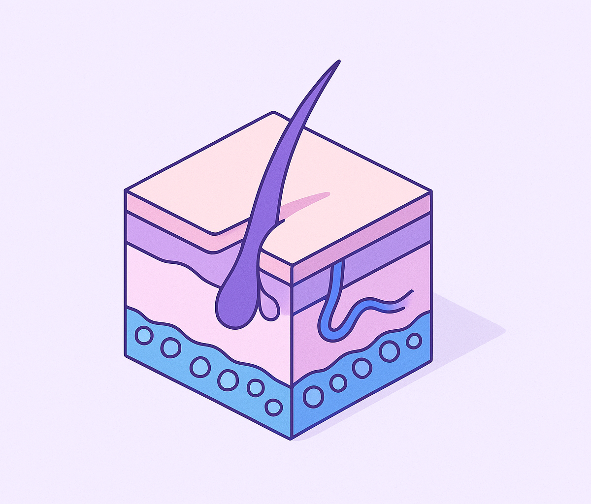
Melanocytes produce melanin, the pigment that absorbs UV radiation and protects skin cells from DNA damage. The relevant player is eumelanin: higher concentrations mean darker phototypes and more built-in UV protection. Darker skin also carries larger, more widely distributed melanosomes and more reactive fibroblasts, structural differences that matter a lot once you're talking about laser treatments or light-based therapies.
More melanin also means photoaging shows up later and less dramatically. That's a genuine biological advantage, but don't read it as immunity. The natural UV protection in very dark skin works out to roughly SPF 13 equivalent. Meaningful, sure, but nowhere near enough to block UV-induced DNA damage on its own.
Lighter skin, on the other end, carries less melanin and a higher share of pheomelanin, which doesn't absorb UV effectively at all. That's the biological root of why melanoma risk skews toward lighter phototypes.
So the biology backs up the scale's basic premise. But it refuses to sort cleanly into six tidy buckets, especially once you're talking about people with mixed ancestry or sun histories that don't follow a single pattern. Skin pigmentation runs on a gradient. The scale runs on steps.
Where the scale genuinely earns its place in clinical practice
None of this means the tool belongs in the trash. Not even close.
Phototherapy is still where the scale does its best work: PUVA, narrowband UVB, exactly the use case it was built for back in 1975. In aesthetics, melanin acts as a chromophore during laser and light-based treatments, absorbing energy and raising the risk of epidermal injury. Phototype guides parameter selection and informed consent, especially for Types IV through VI, where that risk climbs.
Phototype also helps clinicians anticipate postinflammatory hyperpigmentation, keloids, and scarring, all of which turn up more often in higher phototypes. In clinical trials, the scale gets used to check that treatments are tested across a real range of skin types. Per a 2024 JAAD paper, skip that check and trial populations default toward lighter skin without anyone deciding to make it happen.
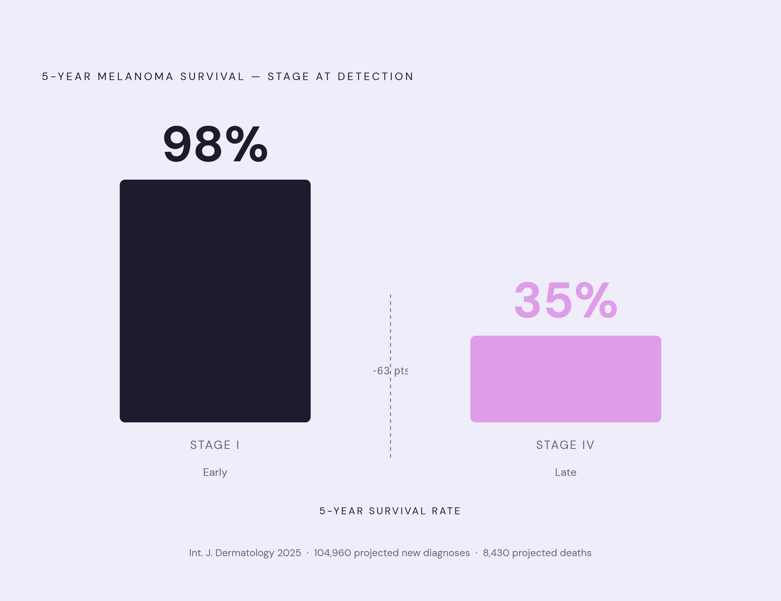
On skin cancer risk, phototype is one input among several, not the whole picture. Dermatologists weigh family history of melanoma, mole count above roughly 50, a history of blistering sunburns (especially in childhood), tanning bed use, immune suppression, and age right alongside it. The stakes for catching things early are enormous: 2025 projections put new melanoma diagnoses at approximately 104,960, with about 8,430 deaths expected. A 2025 study in the International Journal of Dermatology put five-year survival at 98% for Stage I melanoma versus 35% for Stage IV. That gap is why early identification matters so much, and why the scale, imperfect as it is, still earns its keep in the clinic.
Why the scale's original design creates gaps for patients with darker skin tones
So where does it actually break down? Right where its history predicts.
Built entirely on White patients, the scale only started folding in non-white skin types with the 1976 revision, and Types V and VI got defined by skin color alone rather than observed sun reactivity, according to a June 2024 paper in Plastic and Reconstructive Surgery – Global Open. Sit with that for a second: the burn-and-tan logic defining Types I through IV never got applied the same way to V and VI. They were sorted by look, not by behavior. Same scale, different rulebook.
That mismatch shows up at the bedside. The burn-and-tan framework doesn't capture how UV damage actually presents in darker skin. Redness may not be visible, and "burning" may not match what the patient feels, even while real damage happens underneath. Darker skin tones also vary widely in reactivity depending on ethnicity, yet the scale flattens all of that into two categories.
A more recent JAAD paper, from October 2025, put it plainly: the six-category system still doesn't carry enough granularity to represent the full range of darker skin tones in digital and equity-focused research.
The clinical consequence is stark. Roughly 1% of skin cancers occur in Black-identified patients, yet mortality runs two to three times higher for Black-identified individuals compared to White-identified populations, per research indexed in PMC. A big driver is advanced stage at diagnosis. Here's something a lot of practitioners don't know, or don't act on: about 70% of melanomas in skin-of-color individuals show up on the plantar surface of the foot, a site that routine screening often skips entirely.
The categories aren't the only thing failing people here. The vocabulary fails them too. If you don't experience your skin in terms of burning and tanning, you're being asked to find yourself inside a framework that was never built with you in mind.
How self-report and provider assessment introduce further inconsistency
Even setting the historical gaps aside, the process of assigning a type has its own wobble built in.
Patient recall drives a good chunk of the classification, and recall is a loose input by nature. It shifts with sun history, geography, memory, even the season you happen to get asked. Research comparing Fitzpatrick type against objective UV sensitivity testing found the classification wasn't a reliable predictor of measured sensitivity at all. Two people sitting in the same category can respond in completely different ways to sunlight, or to a treatment.
A 2024 study in the Journal of Clinical and Aesthetic Dermatology found something more uncomfortable: a patient's race was a significant factor in discrepancies between how providers and patients described the same person's Fitzpatrick type. That raises real questions about objectivity at the point of care, not just theoretical ones.
Dermatologist assessment, interestingly, turned out to be more accurate than self-report for several of the more-pigmented phototypes. Yet provider assessment isn't bias-free either, and the same 2024 study's authors landed on a fairly stark conclusion: until better stratification methods exist, extreme caution is needed to prevent misuse and overgeneralization of the scale, and patient-guided responses should ultimately determine type.
What does that mean if you're sitting in the exam room? The same person could walk out with different Fitzpatrick classifications from two different providers, or even from the same provider on two different visits. The number on your chart carries a confidence that the process behind it hasn't quite earned.
The problem with using FST as a stand-in for race or skin tone in research
This is where the gaps stack up into something bigger than any single patient visit.
Racial and ethnic data is notoriously hard to record consistently in electronic health records. So researchers reached for something that looked easier to standardize: Fitzpatrick type, used as a proxy for race and ethnicity in dermatology research, according to a January 2024 JAAD paper. Think about what's actually being measured there, though. FST tracks photoreactivity. It says nothing about pigment phenotype, ancestry, or the lived, social experience of race. Swapping one for the other isn't a shortcut, it's a category error.
A paper in the Journal of Investigative Dermatology from August 2025 traced the downstream effect: darker skin phototypes remain underrepresented in clinical trials, and plenty of diagnostic algorithms for skin disease were built using Type I skin as the default reference point.
Follow the loop all the way through, and the shape of the problem gets clearer. The scale gets built on lighter skin. Research then uses that scale as a stand-in for race. Diagnostic tools get trained on the lighter-skin presentations that dominate the resulting data, and patients with darker skin end up less likely to get diagnosed early. That's not a coincidence; it's a documented thread running straight into the outcome disparities from a few sections back.
What patients can actually do with their Fitzpatrick type — and where to press for more
So where does that leave you, sitting across from your own chart with a number next to "Fitzpatrick type"?
Treat it as a starting point for a sun protection conversation, not a verdict. Sunscreen recommendations genuinely differ by type, and it helps to know why. For Types I and II, high-SPF physical blockers built on zinc oxide or titanium dioxide are the standard, since this skin tends to be reactive and burns fast. For higher phototypes, tinted sunscreens with iron oxides often make more sense: they solve the white-cast problem that plain mineral formulas cause on deeper skin, and they add visible-light protection, which matters given the higher risk of hyperpigmentation. Every phototype needs broad-spectrum coverage regardless, since SPF alone handles UVB and sunburn while UVA quietly drives photoaging and skin cancer risk no matter what type you are.
Reject the idea that dark skin is somehow immune to sun damage. It carries lower risk for sunburn and for some cancers, true. Yet postinflammatory pigmentation, scarring tendencies, and later-stage melanoma diagnoses are real risks that don't vanish just because sunburn isn't part of the picture.
A few questions worth bringing to your next visit: is my phototype actually guiding a specific procedure, or is it sitting in my file as a general descriptor nobody's using? If I'm getting a laser treatment or a chemical peel, how does my phototype change the parameters, and what side effects should I watch for? And if you have darker skin, ask directly whether your screening includes less-visible sites like palms, soles, and nail beds, given how often melanoma shows up there and gets missed anyway.
Asynchronous and AI-assisted care can work fine as a first step when something's bothering you about your skin. But the strongest evaluations, whether in person or through a clinician-supervised telehealth visit, weigh the whole picture: phototype alongside personal history, family history, and what your skin actually looks like to a trained eye in the room. Phototype alone was never built to carry that weight by itself, and it still can't.