Hypodermis Function and Its Role in Skin Health
How the deepest skin layer drives aging from beneath the surface.

The hypodermis isn't a fixed, uniform layer sitting quietly under your skin. It shifts constantly, across different body sites, between individuals, and across your own lifespan. That variation is neither random nor purely cosmetic.
Two forces govern it: the number and size of adipocytes in any given region, and hormonal regulation. Testosterone, estrogen, insulin, glucagon, and leptin all direct where fat accumulates and in what quantities. Genetics sets a baseline; hormones revise it continuously, which means the layer you have at forty isn't the layer you had at twenty, and won't be the one you have at sixty.
The sex-based distribution patterns are well-documented. Men accumulate subcutaneous fat in the neck, arms, lower back, and abdomen. Women accumulate it in the breasts, hips, thighs, and buttocks. Neither pattern holds forever. Both shift as hormonal environments change with age, and that shift is precisely why skin appearance changes so predictably across the lifespan. It's not always the surface deteriorating. Sometimes it's the foundation.
Which raises a question worth sitting with: when skin above the hypodermis changes, how often is the real explanation living deeper than the surface where we're looking?
The four core jobs the hypodermis performs for the body and the layers above it
Mechanical cushioning is the most intuitive function. The fat pads of the hypodermis protect muscles and bone from blunt force and sustained pressure. But the connective tissue within this layer also physically tethers skin to underlying structures, maintaining contour. Facial volume loss, then, isn't simply a wrinkle problem. It's a structural one. The scaffold is going.
Thermal insulation is more active than most people expect. The adipose layer slows heat loss, yes, but the hypodermis also participates in temperature regulation through its vasculature. Blood vessels dilate to dissipate heat and constrict to retain it, working in coordination with sweating and piloerection. When the hypodermal layer thins with age, that thermoregulatory buffer weakens. Older adults feel cold more easily, and this is part of the reason, not just reduced circulation in general.
Energy storage is the third function. The triglycerides stored in adipocytes represent long-term metabolic reserve, and the hypodermis responds meaningfully to diet, hormonal fluctuations, and illness. It's a continuous participant in the body's energy economy, not a passive depot.
The fourth function is the one most consequential for skin health: vascular delivery. The dense blood vessel network at the dermis-hypodermis interface supplies oxygen and nutrients upward. The epidermis has no direct blood supply of its own; it relies on diffusion from dermal capillaries, which are themselves fed from below. The hypodermis is the supply chain. Dysfunction at this level has consequences that travel upward through every layer above it, which is why treating only the surface has limits that are baked into the anatomy.
The hypodermis as an endocrine organ and stem cell reservoir: what recent research shows
For a long time, a specific compartment of hypodermal fat called dermal white adipose tissue, or dWAT, was considered functionally similar to other subcutaneous fat. Recent research has clarified that it isn't. The distinction isn't minor.
dWAT secretes adipokines and other signaling molecules that actively influence skin homeostasis. It isn't just storing fat; it's generating biochemical messages that travel upward into the dermis and epidermis. It also holds progenitor cells that participate in skin repair and regeneration. Not peripheral contributors. Essential ones.
A 2024 study published in Communications Biology found that the hypodermis orchestrates the expression of genes critical to skin hydration, development, and differentiation, using full-thickness 3D skin modeling. Read that again slowly: the hypodermis isn't just receiving signals from the layers above it. It's generating signals that regulate what happens above it. Skin health is partly governed from the bottom up.
That finding reframes a persistent frustration in skincare. If you've ever wondered why certain treatments plateau no matter how consistently you apply them, part of the answer is structural. Treatments targeting only the epidermis or dermis operate within limits partly set by the regulatory environment below. Most products simply cannot reach that far.
How hypodermal changes drive the visible signs of skin aging
Collagen and elastin loss are legitimate targets in aging conversations. But some of that degradation originates in the hypodermis, and that distinction matters if you want to understand why some treatments work and others hit a ceiling.
As subcutaneous fat thins with age, a sequence of visible changes unfolds. Sagging and laxity develop because the structural scaffold holding skin taut is diminishing. Facial hollowing at the cheeks and temples, what most people associate with looking older, is volume loss, not wrinkle formation. Fine lines and wrinkles, typically attributed to surface-level changes, are partly downstream of reduced structural support from below. Cold sensitivity increases as the thermoregulatory buffer thins. These aren't isolated phenomena; they're a system degrading at multiple levels simultaneously.
The retinacula cutis, fibrous connective tissue structures beneath the dermis, also contribute to facial contour changes as they weaken. And in aging dWAT specifically, researchers have identified defective adipogenesis, abnormal adipokine secretion, and immune dysfunction. When the hypodermal environment deteriorates, the dermal microenvironment above it deteriorates in parallel.
ECM components including collagen, elastin, and hyaluronic acid are commonly cited as the primary drivers of visible aging. That's not wrong. But the signals accelerating their breakdown don't always originate at the surface. Sometimes they rise from below, and treating the surface alone misses where the process started.
Treatments that work at the hypodermal level: fillers, HA injections, and volume restoration
If the structural deficit causing sagging and contour loss lives in the hypodermis, treating the surface is treating the symptom. The logic for injectable volume restoration follows directly from the anatomy, not from marketing.
Hyaluronic acid is the most widely used filler material. HA is hygroscopic, meaning it binds water and supports tissue hydration and elasticity. A 2025 study found that low-crosslinked HA injected directly into the superficial fat layer showed effectiveness for facial rejuvenation, with high patient satisfaction and no serious adverse events reported.
The limitations are real, though, and worth understanding clearly. Standard HA hydrogels offer rapid volume restoration, but their duration is limited, cell adhesion is weak, and their ability to stimulate collagen regeneration is constrained. Research published in Acta Pharmaceutica Sinica B in 2025 identified the ideal filler profile as combining immediate augmentation, slow degradation, and progressive collagen stimulation. Current HA products partially satisfy that profile. Active research is working to close the gap.
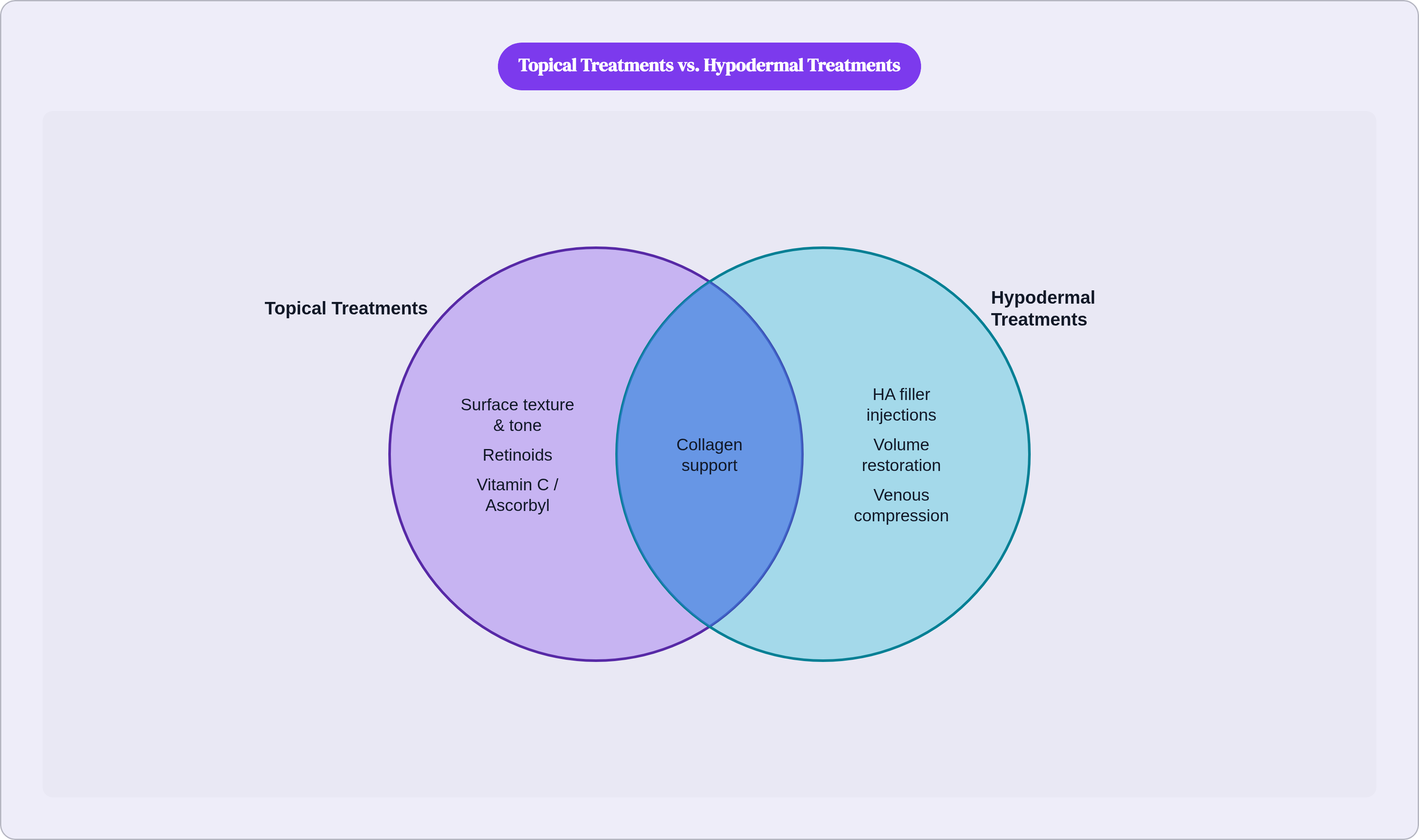
Retinoids stimulate collagen production and inhibit matrix metalloproteinases that degrade existing collagen; those are meaningful, well-documented effects. But when the underlying fat layer has significantly atrophied, topical treatments are working against a structural deficit they cannot resolve. The ceiling on their efficacy is partly set by what lies beneath. Vitamin C in forms like ascorbyl glucoside supports collagen integrity and antioxidant capacity, and it has genuine maintenance value, but it is not a volume restoration tool, and shouldn't be positioned as one.
Topicals address surface texture, tone, and early structural signals. Injectable volume restoration addresses structural deficit at the source. Both have legitimate roles and they are not interchangeable. The confusion between them is where a lot of money gets spent on the wrong solution.
Medical conditions that originate in the hypodermis and how they are diagnosed
Panniculitis is inflammation of subcutaneous fat. It presents as tender, erythematous nodules or plaques, most often on the lower legs, and it covers a heterogeneous group of disorders with overlapping physical findings. Visual assessment alone is insufficient; histopathological correlation, meaning biopsy, is typically required. Management depends on underlying cause and ranges from supportive care to pharmacotherapy to surgical intervention when necrosis or abscess is involved.
Lipodermatosclerosis, or LDS, is a chronic inflammatory condition stemming from longstanding venous insufficiency. It is frequently misdiagnosed as cellulitis because the surface presentation overlaps: pain, redness, swelling. The etiologies are entirely different. Cellulitis is infectious and warrants antibiotics. LDS is inflammatory and warrants compression therapy and venous management. Research published in the British Journal of Nursing in early 2025 centers treatment on reducing venous hypertension. Patients treated for cellulitis when they have LDS receive the wrong treatment, and the underlying condition progresses unchecked. If chronic redness and swelling in the lower legs isn't resolving as expected, it's worth asking whether the diagnosis has actually been confirmed rather than assumed.
Lipodystrophy covers a spectrum of conditions involving dysregulated hypodermal fat: acquired generalized forms, HIV-associated lipodystrophy, drug-induced forms, injection-site lipoatrophy, familial partial lipodystrophy. The consequences extend well beyond appearance. Disrupted glucose metabolism and impaired wound healing are common downstream effects, making this a systemic concern with surface manifestations, not a cosmetic one.
Fourth-degree burns reach the hypodermis, which is what distinguishes them in severity from more superficial burns. Pressure ulcers, abscesses, and tumors can also involve this layer. Debridement, removing necrotic or damaged tissue through ultrasound, laser, pressurized fluid, or surgical means, is a primary treatment modality when the hypodermis is involved in wound pathology.
When a visible skin concern may signal something in the hypodermis worth evaluating
Most everyday texture concerns, dryness, fine lines, dullness, are addressable with evidence-based topical care and involve no hypodermal pathology. That's the baseline, and it's worth holding onto.
What changes the calculus is symptomatic progression. Progressive volume loss or laxity not explained by recent weight change is worth noting. Persistent tender lumps or nodules beneath the skin, particularly on the lower legs, warrant evaluation rather than watchful waiting. Skin changes accompanied by leg swelling or chronic redness should raise the question of LDS before defaulting to an assumption of surface inflammation. Unusual fat redistribution, especially in the context of a new medication or HIV treatment, is a clinical flag.
Tenderness, rapid progression, and systemic symptoms including fatigue or metabolic irregularity are what differentiate a clinical situation from a cosmetic one. When those are present, the next step is evaluation, not a new serum. That distinction sounds obvious stated plainly, but in practice it gets blurred constantly.
Asynchronous teledermatology is a reasonable entry point for photo-documentable concerns. A clinician can often assess from images whether what's visible on the surface warrants investigation at a deeper level.
One thing I've noticed, working through how this layer actually functions: understanding the hypodermis changes how precisely you can describe what you're observing. "Volume loss in the midface" communicates differently than "my skin looks bad." "Tender nodule on the lower leg" lands differently than "redness and swelling." Clearer description produces better clinical conversations. Better clinical conversations produce faster, more accurate diagnoses. That precision isn't a small thing.