Skin Layer Anatomy and Function (New)
Skin's five-layer structure explains how it protects and senses.
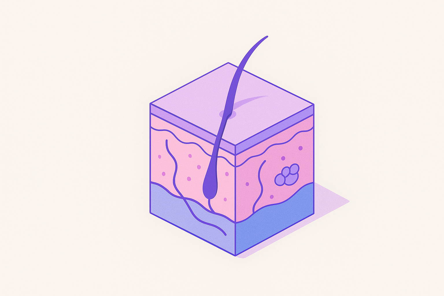
How the Epidermis Is Organized as a Layered Production Line
The skin doesn't get enough credit. We talk about it like it's a wrapper, a surface, something that gets dry in winter. But it covers roughly two square meters, accounts for twelve to fifteen percent of total body weight, and runs five distinct jobs simultaneously: holding water in, keeping pathogens out, regulating temperature, processing sensation, managing immune surveillance. That's a serious organ with a serious architecture.
What makes it particularly interesting isn't any single function. It's that the structure of each layer is essentially the explanation for what it does. Before getting into the layers themselves, one thing worth holding onto: the epidermis, dermis, and hypodermis don't share a developmental origin. The epidermis derives from ectoderm (the outermost embryonic layer); the dermis and hypodermis from mesoderm (the middle embryonic layer). They look structurally unlike each other because they started from entirely different embryological material, and each had to evolve its own toolkit accordingly.
The epidermis itself is keratinized stratified squamous epithelium, layered, toughened skin cells arranged in sheets. No blood vessels of its own. It survives on nutrients diffused upward from the dermis below, which already signals something important about interdependency before we've even gotten started. Keratinocytes make up at least ninety percent of epidermal cells, and their journey from the deepest layer to the surface is the whole organizing logic of the tissue. Cells are born at the base, pushed outward, progressively flattened, chemically transformed, and finally shed as dead, hardened scales. The cycle takes roughly twenty-eight to thirty days. Forty thousand cells shed daily and get replaced.
Thickness varies considerably depending on mechanical demand. On eyelids, the epidermis is about 0.05 mm. On palms and soles, it reaches 1.5 mm. Most skin has four epidermal layers; palms and soles have five, the extra being the stratum lucidum, which appears precisely where friction is highest. None of that variance is arbitrary. But what if the variance itself is the clue? Each structural deviation corresponds to a specific mechanical demand — which means the architecture is not a fixed blueprint but an adaptive response.
What Each Epidermal Layer Does as Keratinocytes Move Outward
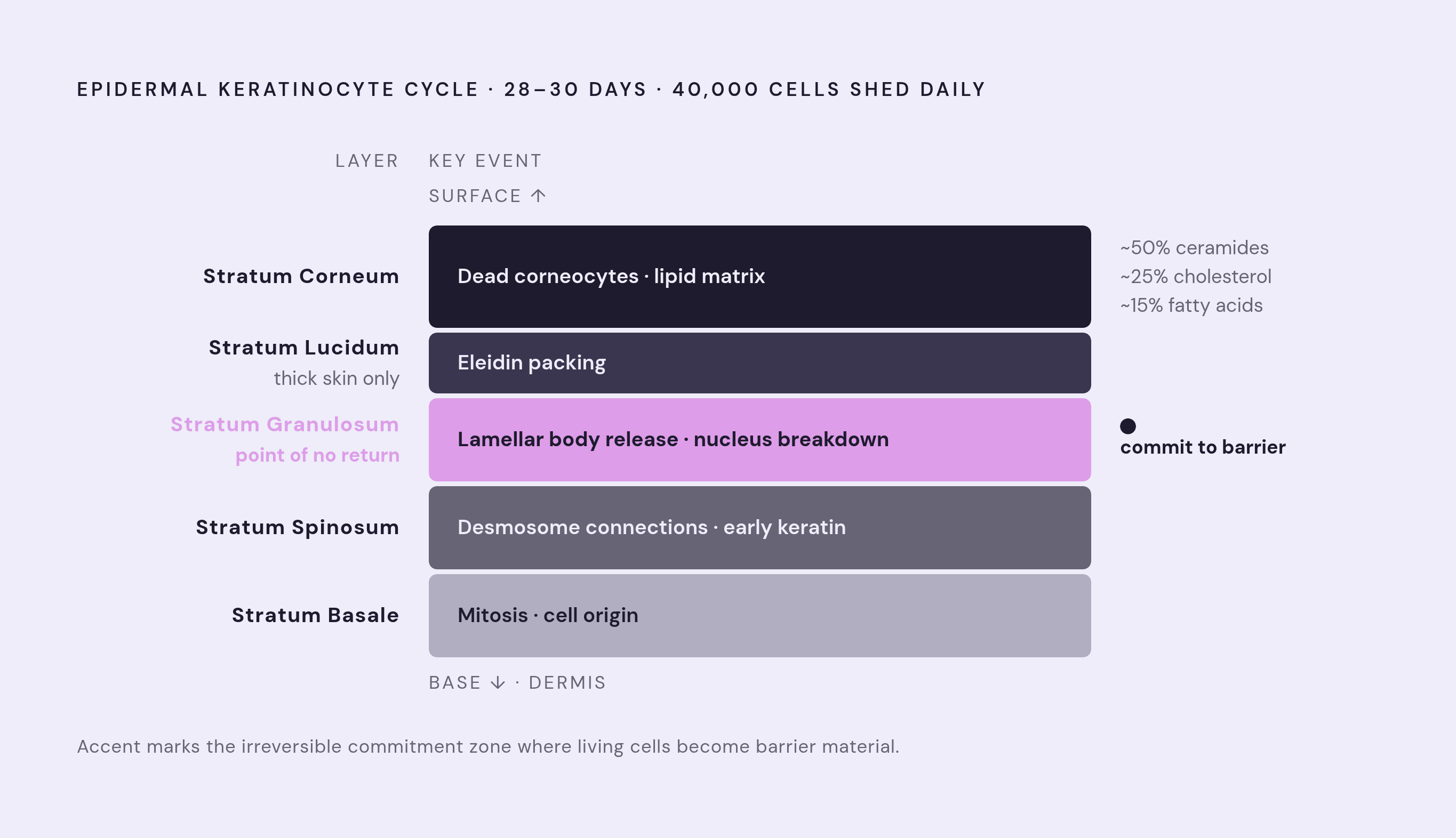
The stratum basale anchors to the basement membrane at the bottom. This is where mitosis happens, where every new keratinocyte originates. Merkel cells also sit here, doing mechanosensory work before the keratinocytes above them have begun their outward journey. The layer is simultaneously a generative zone and a sensory one.
Moving outward, the stratum spinosum is where cells connect via desmosomes, protein structures that look spiky under a microscope. They provide the mechanical cohesion that prevents tissue from shearing apart under lateral force. Early keratin production begins here.
The stratum granulosum is where things become chemically decisive. Cells release lipid-rich lamellar bodies, tiny fat-filled packets, that will form the intercellular lipid matrix above. Nuclei start breaking down. The cell commits, at this point, to becoming barrier material rather than remaining living tissue.
In thick skin only, the stratum lucidum appears just above that, a translucent transitional zone where cells are packed with eleidin, a protein derived from keratin. It's a structural adaptation confined to zones that absorb repeated mechanical stress.
Then the stratum corneum: the finished product. Flattened, nucleus-free dead skin cells (corneocytes) embedded in a lipid matrix composed of roughly fifty percent ceramides, twenty-five percent cholesterol, fifteen percent fatty acids. Corneocytes are the bricks; the lipid matrix is the mortar. What's easy to miss, though, is that the barrier isn't only the stratum corneum. Tight junctions in the living layers below independently restrict outward water movement. That raises an important question: if the stratum corneum is already doing barrier work, why does the skin maintain a second, overlapping restriction system beneath it? Two overlapping systems, the lipid matrix at the surface and tight junctions underneath, both have to be intact for the barrier to hold. Compromise either one, and the other can't fully compensate.
The Three Other Cell Types in the Epidermis and What Each One Contributes
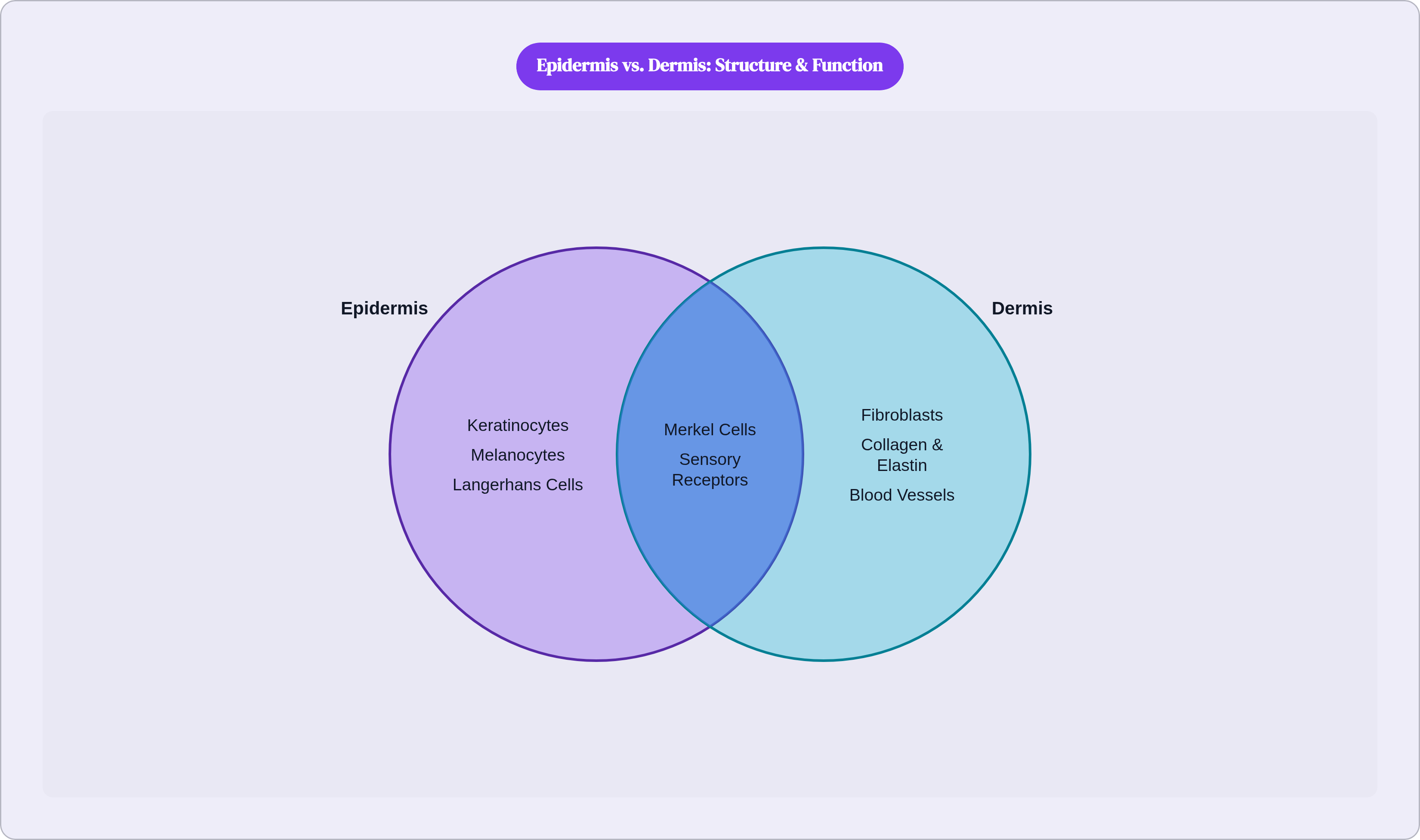
The epidermis isn't only doing barrier work. Three other cell populations are embedded in the same tissue, each running a largely separate operation.
Melanocytes make up roughly eight percent of epidermal cells. They convert tyrosine (an amino acid) into melanin via tyrosinase, an enzyme, and when UVB hits, melanin production ramps up. The granules transfer to neighboring keratinocytes and position over their nuclei like small umbrellas oriented toward the light source. The geometry is deliberate. Melanocytes are doing nuclear protection work, not cosmetic pigmentation.
Langerhans cells are the epidermis's immune agents, derived from bone marrow progenitors, characterized by MHC class II expression (a surface marker that flags them as immune cells) and distinctive Birbeck granules (their hallmark internal structures). When they detect a pathogenic threat, they migrate to lymph nodes to activate a response. But their baseline activity is more interesting than their threat response: under normal conditions, they actively suppress autoinflammation and maintain tolerance toward the skin's resident microorganisms. The immune system here isn't just reactive. It's maintaining a negotiated peace with things it has decided, deliberately, not to fight.
Merkel cells, fewer than one percent of epidermal cells, are pressure-sensing nerve cells (neuroendocrine mechanoreceptors) anchored to keratinocytes by desmosomes, sensitive specifically to sustained light touch. They appear almost redundant given how many sensory structures the dermis carries, until you realize that receptor depth corresponds directly to stimulus profile. Merkel cells detect something that deeper receptors would miss entirely.
So the epidermis is simultaneously building a physical barrier, managing UV exposure, running immune surveillance, and processing light touch, through four distinct cell populations in one tissue. The organizational economy of that doesn't announce itself; you have to stop and look.
How the Dermis Provides the Infrastructure the Epidermis Depends On
The dermis splits into two sublayers. The papillary dermis, sitting just beneath the epidermis, is loose connective tissue, relatively thin. The reticular dermis below it is dense, irregular, well-vascularized, significantly thicker. Between the dermis and the epidermis sits a junction rich in extracellular matrix proteins and growth factors, the structural interface that prevents layer separation and contributes to the skin's overall mechanical integrity.
Fibroblasts are the dermis's dominant cell type. They synthesize and remodel collagen, elastin, glycosaminoglycans (long-chain sugars that attract and hold water), and fibronectin. Two proteins define the dermis's mechanical character in particular. Collagen provides tensile strength and binds water. Elastin enables recoil after deformation. Same tissue, two structurally distinct proteins, two different mechanical jobs. They don't overlap; they complement.
The dermis also carries the blood supply the avascular epidermis cannot generate for itself. Vessels organize into two plexuses: one between the papillary and reticular layers, one at the dermal-hypodermal interface. Nutrients diffuse upward from there. The epidermis has no independent metabolic infrastructure; it borrows everything from the layer beneath it. It is also worth considering what that dependency implies for the epidermis when dermal collagen degrades — the epidermis doesn't just lose structural backing; it loses its nutritional lifeline. That makes the dermis something closer to a life-support system than structural backing, which changes how you think about what happens when that collagen degrades.
Temperature Regulation, Sensation, and the Appendages Housed in the Dermis
The dermal vascular network is the mechanism behind thermoregulation. Increase perfusion and heat moves from the body core toward the environment. Restrict it and heat stays in. That redistribution happens continuously, largely beneath conscious notice, responsive to signals from the central nervous system in ways that are architecturally built into the vasculature itself.
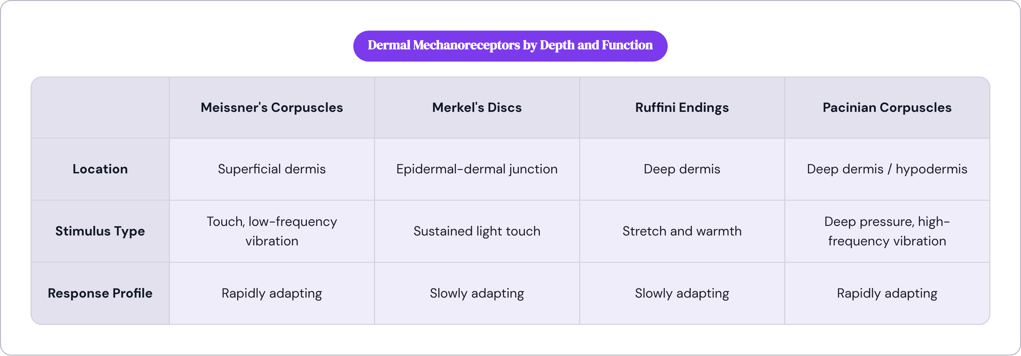
Sensory function in the dermis is organized by the same principle: structure predicts function, and depth predicts stimulus type. Four primary pressure-sensing structures (mechanoreceptors) sit at different depths, each tuned to a different input. Meissner's corpuscles respond to touch and low-frequency vibration. Pacinian corpuscles detect transient pressure and high-frequency vibration. Ruffini endings register stretch and warmth. Meissner's corpuscles and Merkel's discs cluster in the superficial dermis and at the epidermal-dermal junction; Pacinian and Ruffini receptors sit deeper. The placement isn't incidental. It corresponds directly to the mechanical stimulus each receptor is built to resolve, and the information they collectively generate couldn't be produced by any single receptor type or any single depth.
Free nerve endings, the most numerous receptor class, extend up into the mid-epidermis, handling pain, heat, cold, and light touch across the broadest stimulus range of any group.
The dermis also houses hair follicles, sebaceous glands, eccrine and apocrine sweat glands, and lymphatic vessels. These appendages develop embryologically from ectoderm-mesoderm interaction beginning around the tenth to twelfth week of fetal development. Sebaceous glands are small, sac-shaped structures opening into hair follicles, releasing sebum (a natural oil) that protects the hair shaft from brittleness. A small structure solving a specific mechanical problem through its output.
What the Hypodermis Does as the Deepest Layer and How It Connects the Skin to the Rest of the Body
The hypodermis is composed mainly of adipose tissue (fat cells), with sweat glands and Pacinian corpuscles also present. Its mechanical roles are anchoring the dermis to underlying muscle, fascia (the connective tissue wrapping muscles), and bone; cushioning against impact; and insulating against heat loss. Those are real contributions. But calling it passive filler misses what else is happening.
The adipose tissue here stores fat, produces triglycerides (the chemical form fat takes in your blood), and holds vitamin D precursors. There's metabolic activity in the hypodermis with systemic consequences, not just local ones. It's easy to undervalue a layer that doesn't announce itself visually the way the epidermis does, but the hypodermis is doing regulatory work that the layers above it depend on.
The Pacinian corpuscles here detect deep pressure and vibration, extending the sensory range that began with Merkel cells in the stratum basale and continued through the dermis. Sensation across the full thickness of the skin is a distributed array organized by depth and receptor type, and the hypodermis is a functional part of that distribution.
It's also the transition point where skin stops being skin and becomes the structural interface with the musculoskeletal system. Load transfers here. The organ meets everything else here. Less a terminus than a handoff.
How the Skin Microbiome Functions as an Additional Barrier Layer
Something anatomical accounts tend to skip over: the microbiome. The skin hosts trillions of microorganisms, bacteria, archaea, fungi, and viruses, and the Human Microbiome Project established that this community constitutes a recognized microbial barrier layer. Not metaphorically. Functionally.
The interactions are surprisingly concrete. Staphylococcus epidermidis converts sphingomyelin (a fat found in cell membranes) into ceramides, directly supplementing the stratum corneum's lipid matrix. Streptococcus-derived spermidine improves elasticity and reinforces barrier function. Cutibacterium and Corynebacterium produce free fatty acids that maintain the skin's acidic pH. These organisms aren't passengers; they're doing chemical and structural maintenance work the skin's own cells don't accomplish alone.
Which brings Langerhans cells back into the picture. Their immunological tolerance toward the microbiome under baseline conditions isn't passive neglect. It's an actively maintained truce, one the microbial barrier depends on for its continued function. But how does this affect your original picture of the epidermis as a single, self-contained barrier? Disrupt the immune barrier and you compromise the microbial one. Disrupt the microbial one and the immune system escalates responses it was previously calibrated to suppress. Research published in the International Journal of Dermatology and Venereology in March 2024 implicates disruptions to host-microbiota interaction in atopic dermatitis (eczema), psoriasis, and rosacea. The system maintains itself through active negotiation, and when that negotiation fails, the clinical consequences are visible.
Why the Three Layers Only Make Sense as an Integrated System
No single layer is self-sufficient, and the interdependencies aren't incidental to the design. They are the design.
The epidermis has no blood supply. It depends entirely on dermal diffusion for nutrition. The dermis depends on epidermal keratinocytes for surface protection. The hypodermis anchors and buffers both layers from below; without it, external mechanical forces would transmit directly into connective tissue not built to absorb them. Each layer creates conditions the others require.
Sensation runs the full thickness: Merkel cells in the stratum basale, Meissner's and Ruffini endings distributed through the dermis, Pacinian corpuscles in the hypodermis. The sensory system isn't contained in any one zone. It's a distributed array that requires all three layers to function.
The barrier is tiered the same way: stratum corneum lipids, tight junctions in the living layers, Langerhans immune surveillance above that, then the microbial community functioning as a chemical defense layer at the surface. Each tier of protection builds on what sits beneath it, and failure at one level shifts the load carried by the others in ways that aren't always obvious until something goes wrong.
That's what makes the anatomy clinically useful rather than purely academic. A ceramide deficit is a lipid matrix failure. A microbiome disruption is a chemical barrier failure. Collagen loss in the dermis is an infrastructure failure with consequences for everything above it. Each disruption points back to a specific structural role in a specific layer. The three-layer model isn't just descriptive; it's diagnostic. The skin is complicated not because it has three layers, but because those three layers constitute one system, and none of them makes full sense without the others.